 Movie
Movie Controller


+ Open data

- Basic information
| Entry | Database: PDB / ID: 7vh2 | ||||||
|---|---|---|---|---|---|---|---|
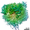
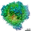
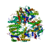
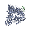
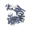
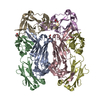
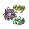
| Title | Cryo-EM structure of Machupo virus dimeric polymerase L | ||||||
 Components Components | RNA-directed RNA polymerase L | ||||||
 Keywords Keywords |  VIRAL PROTEIN / Polymerase VIRAL PROTEIN / Polymerase | ||||||
| Function / homology |  Function and homology information Function and homology informationRNA-templated viral transcription / negative stranded viral RNA replication / cap snatching / virion component / host cell cytoplasm / Hydrolases; Acting on ester bonds / hydrolase activity / RNA-directed RNA polymerase / RNA-dependent RNA polymerase activity / nucleotide binding / metal ion bindingSimilarity search - Function | ||||||
| Biological species |  Machupo virus Machupo virus | ||||||
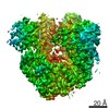
| Method | ELECTRON MICROSCOPY / single particle reconstruction / cryo EM / Resolution: 5.1 Å | ||||||
 Authors Authors | Zhang, X. / Ma, J. / Zhang, S. | ||||||
| Funding support | 1items
| ||||||
 Citation Citation | Journal: Nat Commun / Year: 2021 Title: Structure of Machupo virus polymerase in complex with matrix protein Z. Authors: Jun Ma / Shuangyue Zhang / Xinzheng Zhang /  Abstract: The Arenaviridae family includes several viruses that cause severe human hemorrhagic fevers with high mortality, with no effective countermeasures currently available. The arenavirus multi-domain L ...The Arenaviridae family includes several viruses that cause severe human hemorrhagic fevers with high mortality, with no effective countermeasures currently available. The arenavirus multi-domain L protein is involved in viral transcription and replication and represents a promising target for antiviral drugs. The arenavirus matrix protein Z is a small multi-functional protein that inhibits the activities of the L protein. Here we report the structure of Machupo virus L protein in complex with Z determined by cryo-electron microscopy. The Z protein acts as a staple and binds the L protein with 1:1 stoichiometry at the intersection between the PA-C-like region, RNA-dependent RNA polymerase and PB2-N-like region. Binding of the Z protein may lock the multiple domains of L into a fixed arrangement leading to loss of catalytic activity. These results further our understanding of the inhibitory mechanism of arenavirus replication machinery and provide a novel perspective to develop antiviral drugs. | ||||||
| History |
|
- Structure visualization
| Movie |
Movie viewer |
|---|---|
| Structure viewer | Molecule: MolmilJmol/JSmol |
- Downloads & links
-Download
| PDBx/mmCIF format | 7vh2.cif.gz | 299.2 KB | Display | PDBx/mmCIF format |
|---|---|---|---|---|
| PDB format | pdb7vh2.ent.gz | 240.6 KB | Display | PDB format |
| PDBx/mmJSON format | 7vh2.json.gz | Tree view | PDBx/mmJSON format | |
| Others |  Other downloads Other downloads |
-Validation report
| Arichive directory | https://data.pdbj.org/pub/pdb/validation_reports/vh/7vh2ftp://data.pdbj.org/pub/pdb/validation_reports/vh/7vh2 | HTTPS FTP |
|---|
-Related structure data
| Related structure data | 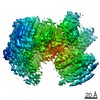 31984MC 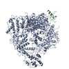 7vgqC  7vh1C  7vh3C C: citing same article ( M: map data used to model this data |
|---|---|
| Similar structure data |


-Links
 PDBj
PDBj- Assembly
| Deposited unit | 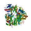
|
|---|---|
| 1 |
|
-Components
| #1: Protein | Mass: 253456.344 Da / Num. of mol.: 1 Source method: isolated from a genetically manipulated source Source: (gene. exp.) Machupo virus / Production host:  Trichoplusia ni (cabbage looper) Trichoplusia ni (cabbage looper)References: UniProt: Q6IUF8, RNA-directed RNA polymerase, Hydrolases; Acting on ester bonds |
|---|
-Experimental details
-Experiment
| Experiment | Method: ELECTRON MICROSCOPY |
|---|---|
| EM experiment | Aggregation state: PARTICLE / 3D reconstruction method: single particle reconstruction |
- Sample preparation
| Component | Name: Machupo virus dimeric polymerase L / Type: COMPLEX / Entity ID: all / Source: RECOMBINANT |
|---|---|
| Source (natural) | Organism: Machupo mammarenavirus |
| Source (recombinant) | Organism: Trichoplusia ni (cabbage looper) |
| Buffer solution | pH: 8 |
| Specimen | Embedding applied: NO / Shadowing applied: NO / Staining applied: NO / Vitrification applied: YES |
| Vitrification | Cryogen name: ETHANE |
- Electron microscopy imaging
| Experimental equipment |  Model: Titan Krios / Image courtesy: FEI Company |
|---|---|
| Microscopy | Model: FEI TITAN KRIOS |
| Electron gun | Electron source: FIELD EMISSION GUN / Accelerating voltage: 300 kV / Illumination mode: FLOOD BEAM |
| Electron lens | Mode: BRIGHT FIELDBright-field microscopy |
| Image recording | Electron dose: 60 e/Å2 / Film or detector model: GATAN K2 SUMMIT (4k x 4k) |
- Processing
| CTF correction | Type: PHASE FLIPPING AND AMPLITUDE CORRECTION |
|---|---|
| 3D reconstruction | Resolution: 5.1 Å / Resolution method: FSC 0.143 CUT-OFF / Num. of particles: 52947 / Symmetry type: POINT |
