 Movie
Movie Controller


+ Open data

- Basic information
| Entry | Database: PDB / ID: 7vd6 | ||||||
|---|---|---|---|---|---|---|---|
| Title | Structure of S1M1-type FCPII complex from diatom | ||||||
 Components Components |
| ||||||
 Keywords Keywords |  ELECTRON TRANSPORT / Photosystem / PSII ELECTRON TRANSPORT / Photosystem / PSII | ||||||
| Function / homology |  Function and homology informationlight-harvesting complex / photosynthesis, light harvesting / plastid / membrane Function and homology informationlight-harvesting complex / photosynthesis, light harvesting / plastid / membraneSimilarity search - Function | ||||||
| Biological species |  Chaetoceros gracilis (Diatom) Chaetoceros gracilis (Diatom) | ||||||
| Method | ELECTRON MICROSCOPY / single particle reconstruction / cryo EM / Resolution: 2.8 Å | ||||||
 Authors Authors | Nagao, R. / Kato, K. / Akita, F. / Miyazaki, N. / Shen, J.R. | ||||||
| Funding support | 1items
| ||||||
 Citation Citation | Journal: Nat Commun / Year: 2022 Title: Structural basis for different types of hetero-tetrameric light-harvesting complexes in a diatom PSII-FCPII supercomplex Authors: Nagao, R. / Kato, K. / Kumazawa, M. / Ifuku, K. / Yokono, M. / Suzuki, T. / Dohmae, N. / Akita, F. / Akimoto, S. / Miyazaki, N. / Shen, J.R. | ||||||
| History |
|
- Structure visualization
| Movie |
Movie viewer |
|---|---|
| Structure viewer | Molecule: MolmilJmol/JSmol |
- Downloads & links
-Download
| PDBx/mmCIF format | 7vd6.cif.gz | 527.6 KB | Display | PDBx/mmCIF format |
|---|---|---|---|---|
| PDB format | pdb7vd6.ent.gz | Display | PDB format | |
| PDBx/mmJSON format | 7vd6.json.gz | Tree view | PDBx/mmJSON format | |
| Others |  Other downloads Other downloads |
-Validation report
| Arichive directory | https://data.pdbj.org/pub/pdb/validation_reports/vd/7vd6ftp://data.pdbj.org/pub/pdb/validation_reports/vd/7vd6 | HTTPS FTP |
|---|
-Related structure data
| Related structure data | 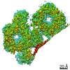 31906MC 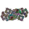 7vd5C M: map data used to model this data C: citing same article ( |
|---|---|
| Similar structure data |


-Links
 PDBj
PDBj


- Assembly
| Deposited unit | 
|
|---|---|
| 1 |
|
-Components
-Protein , 7 types, 11 molecules 1113141518121617192021
| #1: Protein | Mass: 22098.182 Da / Num. of mol.: 5 / Source method: isolated from a natural source / Source: (natural) Chaetoceros gracilis (Diatom) / References: UniProt: A0A679BXP6#2: Protein | | Mass: 22162.188 Da / Num. of mol.: 1 / Source method: isolated from a natural source / Source: (natural) Chaetoceros gracilis (Diatom)#3: Protein | | Mass: 22627.543 Da / Num. of mol.: 1 / Source method: isolated from a natural source / Source: (natural) Chaetoceros gracilis (Diatom)#4: Protein | | Mass: 22298.115 Da / Num. of mol.: 1 / Source method: isolated from a natural source / Source: (natural) Chaetoceros gracilis (Diatom)#5: Protein | | Mass: 29014.037 Da / Num. of mol.: 1 / Source method: isolated from a natural source / Source: (natural) Chaetoceros gracilis (Diatom)#6: Protein | | Mass: 23934.375 Da / Num. of mol.: 1 / Source method: isolated from a natural source / Source: (natural) Chaetoceros gracilis (Diatom)#7: Protein | | Mass: 21143.453 Da / Num. of mol.: 1 / Source method: isolated from a natural source / Source: (natural) Chaetoceros gracilis (Diatom) |
|---|
-Sugars , 1 types, 1 molecules 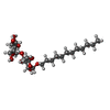
| #16: Sugar | ChemComp-LMU /  Type: D-saccharide / Mass: 510.615 Da / Num. of mol.: 1 / Source method: obtained synthetically / Formula: C24H46O11 / Comment: detergent*YM Type: D-saccharide / Mass: 510.615 Da / Num. of mol.: 1 / Source method: obtained synthetically / Formula: C24H46O11 / Comment: detergent*YM |
|---|
-Non-polymers , 9 types, 208 molecules 
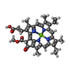


| #8: Chemical | ChemComp-CLA / Chlorophyll a Mass: 893.489 Da / Num. of mol.: 71 / Source method: obtained synthetically / Formula: C55H72MgN4O5 Mass: 893.489 Da / Num. of mol.: 71 / Source method: obtained synthetically / Formula: C55H72MgN4O5#9: Chemical | ChemComp-KC1 / Chlorophyll c Mass: 610.941 Da / Num. of mol.: 35 / Source method: obtained synthetically / Formula: C35H30MgN4O5 Mass: 610.941 Da / Num. of mol.: 35 / Source method: obtained synthetically / Formula: C35H30MgN4O5#10: Chemical | ChemComp-A86 / ( Fucoxanthin Mass: 658.906 Da / Num. of mol.: 54 / Source method: obtained synthetically / Formula: C42H58O6 Mass: 658.906 Da / Num. of mol.: 54 / Source method: obtained synthetically / Formula: C42H58O6#11: Chemical | ChemComp-LMG /  Mass: 787.158 Da / Num. of mol.: 4 / Source method: obtained synthetically / Formula: C45H86O10 Mass: 787.158 Da / Num. of mol.: 4 / Source method: obtained synthetically / Formula: C45H86O10#12: Chemical | ChemComp-UNL / Num. of mol.: 19 / Source method: obtained synthetically #13: Chemical | ChemComp-DD6 / ( Diadinoxanthin Mass: 582.855 Da / Num. of mol.: 6 / Source method: obtained synthetically / Formula: C40H54O3 Mass: 582.855 Da / Num. of mol.: 6 / Source method: obtained synthetically / Formula: C40H54O3#14: Chemical |  Mass: 795.116 Da / Num. of mol.: 2 / Source method: obtained synthetically / Formula: C41H78O12S Mass: 795.116 Da / Num. of mol.: 2 / Source method: obtained synthetically / Formula: C41H78O12S#15: Chemical | ChemComp-LHG / Phosphatidylglycerol Mass: 722.970 Da / Num. of mol.: 4 / Source method: obtained synthetically / Formula: C38H75O10P / Comment: phospholipid*YM Mass: 722.970 Da / Num. of mol.: 4 / Source method: obtained synthetically / Formula: C38H75O10P / Comment: phospholipid*YM#17: Water | ChemComp-HOH / | WaterMass: 18.015 Da / Num. of mol.: 13 / Source method: isolated from a natural source / Formula: H2O |
|---|
-Details
| Has ligand of interest | N |
|---|
-Experimental details
-Experiment
| Experiment | Method: ELECTRON MICROSCOPY |
|---|---|
| EM experiment | Aggregation state: PARTICLE / 3D reconstruction method: single particle reconstruction |
- Sample preparation
| Component | Name: S1M1-type FCPII complex / Type: COMPLEX / Entity ID: #1-#7 / Source: NATURAL | ||||||||||||
|---|---|---|---|---|---|---|---|---|---|---|---|---|---|
| Molecular weight | Value: 0.24 MDa / Experimental value: NO | ||||||||||||
| Source (natural) | Organism: Chaetoceros gracilis (Diatom) | ||||||||||||
| Buffer solution | pH: 6.5 | ||||||||||||
| Buffer component |
| ||||||||||||
| Specimen | Conc.: 0.256 mg/ml / Embedding applied: NO / Shadowing applied: NO / Staining applied: NO / Vitrification applied: YES | ||||||||||||
| Specimen support | Grid material: COPPER / Grid mesh size: 200 divisions/in. / Grid type: Quantifoil R1.2/1.3 | ||||||||||||
| Vitrification | Instrument: FEI VITROBOT MARK IV / Cryogen name: ETHANE / Humidity: 100 % / Chamber temperature: 277 K |
- Electron microscopy imaging
| Experimental equipment |  Model: Titan Krios / Image courtesy: FEI Company |
|---|---|
| Microscopy | Model: FEI TITAN KRIOS |
| Electron gun | Electron source: FIELD EMISSION GUN / Accelerating voltage: 300 kV / Illumination mode: FLOOD BEAM |
| Electron lens | Mode: BRIGHT FIELDBright-field microscopy |
| Image recording | Electron dose: 20 e/Å2 / Film or detector model: FEI FALCON III (4k x 4k) |
- Processing
| EM software |
| ||||||||||||||||||||||||||||||||||||
|---|---|---|---|---|---|---|---|---|---|---|---|---|---|---|---|---|---|---|---|---|---|---|---|---|---|---|---|---|---|---|---|---|---|---|---|---|---|
| CTF correction | Type: PHASE FLIPPING AND AMPLITUDE CORRECTION | ||||||||||||||||||||||||||||||||||||
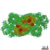
| Particle selection | Num. of particles selected: 8093924 | ||||||||||||||||||||||||||||||||||||
| Symmetry | Point symmetry: C1 (asymmetric) | ||||||||||||||||||||||||||||||||||||
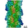
| 3D reconstruction | Resolution: 2.8 Å / Resolution method: FSC 0.143 CUT-OFF / Num. of particles: 373897 / Algorithm: FOURIER SPACE / Symmetry type: POINT | ||||||||||||||||||||||||||||||||||||
| Atomic model building | Protocol: FLEXIBLE FIT / Space: REAL / Target criteria: Correlation coefficient | ||||||||||||||||||||||||||||||||||||
| Atomic model building | PDB-ID: 6J40 |

