 Movie
Movie Controller


+ Open data

- Basic information
| Entry | Database: PDB / ID: 7v8h | ||||||||||||||||||
|---|---|---|---|---|---|---|---|---|---|---|---|---|---|---|---|---|---|---|---|
| Title | Crystal structure of LRR domain from Shigella flexneri IpaH1.4 | ||||||||||||||||||
 Components Components | RING-type E3 ubiquitin transferase | ||||||||||||||||||
 Keywords Keywords |  LIGASE / ubiquitin / innate immune LIGASE / ubiquitin / innate immune | ||||||||||||||||||
| Function / homology |  Function and homology information Function and homology informationmodulation of process of another organism / RING-type E3 ubiquitin transferase / ubiquitin-protein transferase activity / host cell cytoplasm / protein ubiquitination / extracellular region / cytoplasmSimilarity search - Function | ||||||||||||||||||
| Biological species |  Shigella flexneri 5a str. M90T (bacteria) Shigella flexneri 5a str. M90T (bacteria) | ||||||||||||||||||
| Method | X-RAY DIFFRACTION / SYNCHROTRON / MOLECULAR REPLACEMENT / Resolution: 2.46 Å | ||||||||||||||||||
 Authors Authors | Liu, J. / Wang, Y. / Pan, L. | ||||||||||||||||||
| Funding support |  China, 5items China, 5items
| ||||||||||||||||||
 Citation Citation | Journal: Proc.Natl.Acad.Sci.USA / Year: 2022 Title: Mechanistic insights into the subversion of the linear ubiquitin chain assembly complex by the E3 ligase IpaH1.4 of Shigella flexneri. Authors: Liu, J. / Wang, Y. / Wang, D. / Wang, Y. / Xu, X. / Zhang, Y. / Li, Y. / Zhang, M. / Gong, X. / Tang, Y. / Shen, L. / Li, M. / Pan, L. | ||||||||||||||||||
| History |
|
- Structure visualization
| Structure viewer | Molecule: MolmilJmol/JSmol |
|---|
- Downloads & links
-Download
| PDBx/mmCIF format | 7v8h.cif.gz | 127.7 KB | Display | PDBx/mmCIF format |
|---|---|---|---|---|
| PDB format | pdb7v8h.ent.gz | 79.7 KB | Display | PDB format |
| PDBx/mmJSON format | 7v8h.json.gz | Tree view | PDBx/mmJSON format | |
| Others |  Other downloads Other downloads |
-Validation report
| Arichive directory | https://data.pdbj.org/pub/pdb/validation_reports/v8/7v8hftp://data.pdbj.org/pub/pdb/validation_reports/v8/7v8h | HTTPS FTP |
|---|
-Related structure data
| Related structure data | 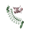 7v8eC  7v8fC 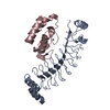 7v8gC 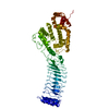 5kh1S S: Starting model for refinement C: citing same article ( |
|---|---|
| Similar structure data |
-Links
 PDBj
PDBj


- Assembly
| Deposited unit | 
| ||||||||||||||||||||||||||||||||||||||||||||
|---|---|---|---|---|---|---|---|---|---|---|---|---|---|---|---|---|---|---|---|---|---|---|---|---|---|---|---|---|---|---|---|---|---|---|---|---|---|---|---|---|---|---|---|---|---|
| 1 |
| ||||||||||||||||||||||||||||||||||||||||||||
| Unit cell |
| ||||||||||||||||||||||||||||||||||||||||||||
| Noncrystallographic symmetry (NCS) | NCS domain:
NCS domain segments:
NCS oper: (Code: givenMatrix: (-0.909535870212, -0.0948495899864, 0.40465794948), (-0.0753447108192, -0.919853403415, -0.384958297452), (0.408739128723, -0.380622216253, 0.829493250814)Vector: 62. ...NCS oper: (Code: given Matrix: (-0.909535870212, -0.0948495899864, 0.40465794948), Vector : |
-Components
| #1: Protein | Mass: 26991.988 Da / Num. of mol.: 2 Source method: isolated from a genetically manipulated source Source: (gene. exp.) Shigella flexneri 5a str. M90T (bacteria)Strain: M90T / Gene: ipaH1.4 / Plasmid: pRSF-Deut1-Trx / Production host: Escherichia coli BL21(DE3) (bacteria) / Strain (production host): BL21(DE3)References: UniProt: Q9AFJ5, RING-type E3 ubiquitin transferase #2: Water | ChemComp-HOH / | Water Mass: 18.015 Da / Num. of mol.: 90 / Source method: isolated from a natural source / Formula: H2O Mass: 18.015 Da / Num. of mol.: 90 / Source method: isolated from a natural source / Formula: H2O |
|---|
-Experimental details
-Experiment
| Experiment | Method: X-RAY DIFFRACTION / Number of used crystals: 1 |
|---|
- Sample preparation
| Crystal | Density Matthews: 2.75 Å3/Da / Density % sol: 55.31 % |
|---|---|
| Crystal grow | Temperature: 289 K / Method: evaporation / pH: 4 / Details: 0.1 M sodium citrate (pH 4.0), 0.8 M (NH4)2SO4 |
-Data collection
| Diffraction | Mean temperature: 100 K / Serial crystal experiment: N |
|---|---|
| Diffraction source | Source: SYNCHROTRON / Site: SSRF / Beamline: BL19U1 / Wavelength: 0.9793 Å |
| Detector | Type: DECTRIS PILATUS 6M / Detector: PIXEL / Date: Nov 3, 2018 |
| Radiation | Protocol: SINGLE WAVELENGTH / Monochromatic (M) / Laue (L): M / Scattering type: x-ray |
| Radiation wavelength | Wavelength: 0.9793 Å / Relative weight: 1 |
| Reflection | Resolution: 2.456→76.919 Å / Num. obs: 21136 / % possible obs: 99.8 % / Observed criterion σ(I): 2 / Redundancy: 24.6 % / Biso Wilson estimate: 37.05 Å2 / CC1/2: 0.998 / Rpim(I) all: 0.067 / Net I/σ(I): 14.4 |
| Reflection shell | Resolution: 2.456→2.498 Å / Redundancy: 25.4 % / Mean I/σ(I) obs: 3.1 / Num. unique obs: 1091 / CC1/2: 0.81 / Rpim(I) all: 0.518 / % possible all: 100 |
- Processing
| Software |
| |||||||||||||||||||||||||||||||||||||||||||||||||||||||||||||||
|---|---|---|---|---|---|---|---|---|---|---|---|---|---|---|---|---|---|---|---|---|---|---|---|---|---|---|---|---|---|---|---|---|---|---|---|---|---|---|---|---|---|---|---|---|---|---|---|---|---|---|---|---|---|---|---|---|---|---|---|---|---|---|---|---|
| Refinement | Method to determine structure: MOLECULAR REPLACEMENT Starting model: 5KH1 Resolution: 2.46→60.82 Å / SU ML: 0.2194 / Cross valid method: FREE R-VALUE / σ(F): 1.34 / Phase error: 22.7769 Stereochemistry target values: GeoStd + Monomer Library + CDL v1.2
| |||||||||||||||||||||||||||||||||||||||||||||||||||||||||||||||
| Solvent computation | Shrinkage radii: 0.9 Å / VDW probe radii: 1.11 Å / Solvent model: FLAT BULK SOLVENT MODEL | |||||||||||||||||||||||||||||||||||||||||||||||||||||||||||||||
| Displacement parameters | Biso mean: 39.53 Å2 | |||||||||||||||||||||||||||||||||||||||||||||||||||||||||||||||
| Refinement step | Cycle: LAST / Resolution: 2.46→60.82 Å
| |||||||||||||||||||||||||||||||||||||||||||||||||||||||||||||||
| Refine LS restraints |
| |||||||||||||||||||||||||||||||||||||||||||||||||||||||||||||||
| Refine LS restraints NCS | Type: Torsion NCS / Rms dev position: 0.734799407111 Å | |||||||||||||||||||||||||||||||||||||||||||||||||||||||||||||||
| LS refinement shell |
|
