 Movie
Movie Controller


+ Open data

- Basic information
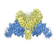
| Entry | Database: PDB / ID: 7v7c | ||||||||||||||||||
|---|---|---|---|---|---|---|---|---|---|---|---|---|---|---|---|---|---|---|---|
| Title | CryoEM structure of DDB1-VprBP-Vpr-UNG2(94-313) complex | ||||||||||||||||||
 Components Components |
| ||||||||||||||||||
 Keywords Keywords | TRANSFERASE/DNA BINDING PROTEIN/VIRAL PROTEIN /  E3 ligase / HIV accessory protein / restriction factors / LIGASE / TRANSFERASE-DNA BINDING PROTEIN-VIRAL PROTEIN complex E3 ligase / HIV accessory protein / restriction factors / LIGASE / TRANSFERASE-DNA BINDING PROTEIN-VIRAL PROTEIN complex | ||||||||||||||||||
| Function / homology |  Function and homology information Function and homology informationhistone H2AT120 kinase activity / cell competition in a multicellular organism / symbiont-mediated arrest of host cell cycle during G2/M transition / base-excision repair, AP site formation via deaminated base removal / uracil-DNA glycosylase / depyrimidination / Displacement of DNA glycosylase by APEX1 / positive regulation by virus of viral protein levels in host cell / V(D)J recombination / isotype switching ...histone H2AT120 kinase activity / cell competition in a multicellular organism / symbiont-mediated arrest of host cell cycle during G2/M transition / base-excision repair, AP site formation via deaminated base removal / uracil-DNA glycosylase / depyrimidination / Displacement of DNA glycosylase by APEX1 / positive regulation by virus of viral protein levels in host cell / V(D)J recombination / isotype switching / epigenetic programming in the zygotic pronuclei / spindle assembly involved in female meiosis / Cul4-RING E3 ubiquitin ligase complex / UV-damage excision repair / uracil DNA N-glycosylase activity / biological process involved in interaction with symbiont / regulation of mitotic cell cycle phase transition / WD40-repeat domain binding / Cul4A-RING E3 ubiquitin ligase complex / Cul4B-RING E3 ubiquitin ligase complex / ubiquitin ligase complex scaffold activity / negative regulation of reproductive process / negative regulation of developmental process / cullin family protein binding / ribosomal small subunit binding / ubiquitin-like ligase-substrate adaptor activity / viral release from host cell / ectopic germ cell programmed cell death / somatic hypermutation of immunoglobulin genes / positive regulation of viral genome replication / monoatomic ion transport / positive regulation of gluconeogenesis / Recognition and association of DNA glycosylase with site containing an affected pyrimidine / Cleavage of the damaged pyrimidine / post-translational protein modification / B cell differentiation / Chromatin modifications during the maternal to zygotic transition (MZT) / virion component / proteasomal protein catabolic process / nuclear estrogen receptor binding / nucleotide-excision repair / Recognition of DNA damage by PCNA-containing replication complex / DNA Damage Recognition in GG-NER / base-excision repair / : / protein homooligomerization / regulation of circadian rhythm / Dual Incision in GG-NER / Transcription-Coupled Nucleotide Excision Repair (TC-NER) / Formation of TC-NER Pre-Incision Complex / fibrillar center / Wnt signaling pathway / Formation of Incision Complex in GG-NER / Dual incision in TC-NER / Gap-filling DNA repair synthesis and ligation in TC-NER / viral penetration into host nucleus / positive regulation of protein catabolic process / cellular response to UV / rhythmic process / protein-macromolecule adaptor activity / Antigen processing: Ubiquitination & Proteasome degradation / site of double-strand break / Neddylation / ubiquitin-dependent protein catabolic process / proteasome-mediated ubiquitin-dependent protein catabolic process / host extracellular space / chromosome, telomeric region / damaged DNA binding / protein ubiquitination / non-specific serine/threonine protein kinase / symbiont entry into host cell / cell cycle / phosphorylation / protein serine kinase activity / DNA repair / DNA-templated transcription / apoptotic process / DNA damage response / host cell nucleus / protein-containing complex binding / nucleolus / regulation of DNA-templated transcription / negative regulation of apoptotic process / negative regulation of transcription by RNA polymerase II / protein-containing complex / mitochondrion / DNA binding / extracellular space / extracellular exosome / nucleoplasm / ATP binding / nucleus / cytoplasmSimilarity search - Function | ||||||||||||||||||
| Biological species |  Homo sapiens (human) Homo sapiens (human)  Human immunodeficiency virus 1 Human immunodeficiency virus 1 | ||||||||||||||||||
| Method | ELECTRON MICROSCOPY / single particle reconstruction / cryo EM / Resolution: 3.7 Å | ||||||||||||||||||
 Authors Authors | Wang, D. / Xu, J. / Liu, Q. / Xiang, Y. | ||||||||||||||||||
| Funding support |  China, 5items China, 5items
| ||||||||||||||||||
 Citation Citation | Journal: To Be Published Title: Structural insights into the HIV-1 Vpr mediated ubiquitination through the Cullin-RING E3 ubiquitin ligase Authors: Wang, D. / Xu, J. / Liu, Q. / Xiang, Y. | ||||||||||||||||||
| History |
|
- Structure visualization
| Structure viewer | Molecule: MolmilJmol/JSmol |
|---|
- Downloads & links
-Download
| PDBx/mmCIF format | 7v7c.cif.gz | 882.7 KB | Display | PDBx/mmCIF format |
|---|---|---|---|---|
| PDB format | pdb7v7c.ent.gz | 720.2 KB | Display | PDB format |
| PDBx/mmJSON format | 7v7c.json.gz | Tree view | PDBx/mmJSON format | |
| Others |  Other downloads Other downloads |
-Validation report
| Arichive directory | https://data.pdbj.org/pub/pdb/validation_reports/v7/7v7cftp://data.pdbj.org/pub/pdb/validation_reports/v7/7v7c | HTTPS FTP |
|---|
-Related structure data
| Related structure data |  31766MC 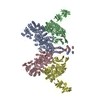 7v7bC M: map data used to model this data C: citing same article ( |
|---|---|
| Similar structure data |
-Links
 PDBj
PDBj


- Assembly
| Deposited unit | 
|
|---|---|
| 1 |
|
-Components
| #1: Protein | Mass: 169194.781 Da / Num. of mol.: 2 Source method: isolated from a genetically manipulated source Source: (gene. exp.) Homo sapiens (human) / Gene: DCAF1, KIAA0800, RIP, VPRBP / Production host:  Trichoplusia ni (cabbage looper) Trichoplusia ni (cabbage looper)References: UniProt: Q9Y4B6, non-specific serine/threonine protein kinase#2: Protein | Mass: 127097.469 Da / Num. of mol.: 2 Source method: isolated from a genetically manipulated source Source: (gene. exp.) Homo sapiens (human) / Gene: DDB1, XAP1 / Production host: Trichoplusia ni (cabbage looper) / References: UniProt: Q16531#3: Protein | Mass: 11396.878 Da / Num. of mol.: 2 Source method: isolated from a genetically manipulated source Source: (gene. exp.) Human immunodeficiency virus 1 / Gene: vpr / Production host:  Escherichia coli (E. coli) / References: UniProt: B2CPZ1 Escherichia coli (E. coli) / References: UniProt: B2CPZ1#4: Protein | / UDGMass: 25136.654 Da / Num. of mol.: 2 Source method: isolated from a genetically manipulated source Source: (gene. exp.) Homo sapiens (human) / Gene: UNG, DGU, UNG1, UNG15 / Production host: Escherichia coli (E. coli) / References: UniProt: P13051, uracil-DNA glycosylase |
|---|
-Experimental details
-Experiment
| Experiment | Method: ELECTRON MICROSCOPY |
|---|---|
| EM experiment | Aggregation state: PARTICLE / 3D reconstruction method: single particle reconstruction |
- Sample preparation
| Component | Name: DDB1-VprBP-Vpr-UNG2(94-313) complex / Type: COMPLEX / Entity ID: all / Source: RECOMBINANT |
|---|---|
| Molecular weight | Value: 0.66 kDa/nm / Experimental value: NO |
| Source (natural) | Organism: Homo sapiens (human) |
| Source (recombinant) | Organism: Trichoplusia ni (cabbage looper) |
| Buffer solution | pH: 7.5 |
| Specimen | Embedding applied: NO / Shadowing applied: NO / Staining applied: NO / Vitrification applied: YES |
| Vitrification | Cryogen name: ETHANE |
- Electron microscopy imaging
| Experimental equipment |  Model: Titan Krios / Image courtesy: FEI Company |
|---|---|
| Microscopy | Model: FEI TITAN KRIOS |
| Electron gun | Electron source: FIELD EMISSION GUN / Accelerating voltage: 300 kV / Illumination mode: FLOOD BEAM |
| Electron lens | Mode: BRIGHT FIELDBright-field microscopy |
| Image recording | Electron dose: 50 e/Å2 / Film or detector model: GATAN K3 (6k x 4k) |
- Processing
| Software | Name: PHENIX / Version: 1.13_2998: / Classification: refinement | |||||||||||||||||||||
|---|---|---|---|---|---|---|---|---|---|---|---|---|---|---|---|---|---|---|---|---|---|---|
| EM software |
| |||||||||||||||||||||
| CTF correction | Type: NONE | |||||||||||||||||||||
| 3D reconstruction | Resolution: 3.7 Å / Resolution method: FSC 0.143 CUT-OFF / Num. of particles: 145568 / Symmetry type: POINT | |||||||||||||||||||||
| Atomic model building | Protocol: RIGID BODY FIT |
