 Movie
Movie Controller

[English] 日本語
 Yorodumi
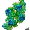
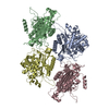
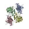
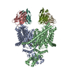
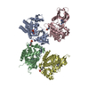
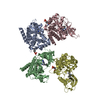
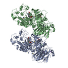
- PDB-7v1y: Serine beta-lactamase-like protein LACTB in complex with inhibitor -
+ Open data

- Basic information
| Entry | Database: PDB / ID: 7v1y | |||||||||
|---|---|---|---|---|---|---|---|---|---|---|
| Title | Serine beta-lactamase-like protein LACTB in complex with inhibitor | |||||||||
 Components Components |
| |||||||||
 Keywords Keywords |  HYDROLASE / mitochondrial intermembrane space protease / CYTOSOLIC PROTEIN HYDROLASE / mitochondrial intermembrane space protease / CYTOSOLIC PROTEIN | |||||||||
| Function / homology |  Function and homology informationHydrolases; Acting on peptide bonds (peptidases) / regulation of lipid metabolic process / lipid metabolic process / peptidase activity / mitochondrion / proteolysis / identical protein binding / cytosol Function and homology informationHydrolases; Acting on peptide bonds (peptidases) / regulation of lipid metabolic process / lipid metabolic process / peptidase activity / mitochondrion / proteolysis / identical protein binding / cytosolSimilarity search - Function | |||||||||
| Biological species |  Homo sapiens (human) Homo sapiens (human)synthetic construct (others) | |||||||||
| Method | ELECTRON MICROSCOPY / single particle reconstruction / cryo EM / Resolution: 2.82 Å | |||||||||
 Authors Authors | Zhang, M.H. / Yang, M.J. | |||||||||
| Funding support |  China, 1items China, 1items
| |||||||||
 Citation Citation | Journal: Structure / Year: 2022 Title: Structural basis for the catalytic activity of filamentous human serine beta-lactamase-like protein LACTB. Authors: Minghui Zhang / Laixing Zhang / Runyu Guo / Chun Xiao / Jian Yin / Sensen Zhang / Maojun Yang / Abstract: Serine beta-lactamase-like protein (LACTB) is a mammalian mitochondrial serine protease that can specifically hydrolyze peptide bonds adjacent to aspartic acid residues and is structurally related to ...Serine beta-lactamase-like protein (LACTB) is a mammalian mitochondrial serine protease that can specifically hydrolyze peptide bonds adjacent to aspartic acid residues and is structurally related to prokaryotic penicillin-binding proteins. Here, we determined the cryoelectron microscopy structures of human LACTB (hLACTB) filaments from wild-type protein, a middle region deletion mutant, and in complex with the inhibitor Z-AAD-CMK at 3.0-, 3.1-, and 2.8-Å resolution, respectively. Structural analysis and activity assays revealed that three interfaces are required for the assembly of hLACTB filaments and that the formation of higher order helical structures facilitates its cleavage activity. Further structural and enzymatic analyses of middle region deletion constructs indicated that, while this region is necessary for substrate hydrolysis, it is not required for filament formation. Moreover, the inhibitor-bound structure showed that hLACTB may cleave peptide bonds adjacent to aspartic acid residues. These findings provide the structural basis underlying hLACTB catalytic activity. | |||||||||
| History |
|
- Structure visualization
| Movie |
Movie viewer |
|---|---|
| Structure viewer | Molecule: MolmilJmol/JSmol |
- Downloads & links
-Download
| PDBx/mmCIF format | 7v1y.cif.gz | 282.3 KB | Display | PDBx/mmCIF format |
|---|---|---|---|---|
| PDB format | pdb7v1y.ent.gz | 228.3 KB | Display | PDB format |
| PDBx/mmJSON format | 7v1y.json.gz | Tree view | PDBx/mmJSON format | |
| Others |  Other downloads Other downloads |
-Validation report
| Arichive directory | https://data.pdbj.org/pub/pdb/validation_reports/v1/7v1yftp://data.pdbj.org/pub/pdb/validation_reports/v1/7v1y | HTTPS FTP |
|---|
-Related structure data
| Related structure data | 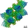 31630MC 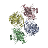 7v1zC 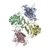 7v21C M: map data used to model this data C: citing same article ( |
|---|---|
| Similar structure data |


-Links
 PDBj
PDBj
- Assembly
| Deposited unit | 
|
|---|---|
| 1 |
|
-Components
| #1: Protein | Mass: 54889.445 Da / Num. of mol.: 4 Source method: isolated from a genetically manipulated source Source: (gene. exp.) Homo sapiens (human) / Gene: LACTB, MRPL56, UNQ843/PRO1781 / Production host:  Escherichia coli (E. coli) Escherichia coli (E. coli)References: UniProt: P83111, Hydrolases; Acting on peptide bonds (peptidases)#2: Protein/peptide | Mass: 261.275 Da / Num. of mol.: 4 / Source method: obtained synthetically / Source: (synth.) synthetic construct (others) Has ligand of interest | Y | |
|---|
-Experimental details
-Experiment
| Experiment | Method: ELECTRON MICROSCOPY |
|---|---|
| EM experiment | Aggregation state: PARTICLE / 3D reconstruction method: single particle reconstruction |
- Sample preparation
| Component | Name: Mitochondrial intermembrane space protease. / Type: ORGANELLE OR CELLULAR COMPONENT / Entity ID: #1 / Source: RECOMBINANT |
|---|---|
| Molecular weight | Value: 55 kDa/nm / Experimental value: NO |
| Source (natural) | Organism: Homo sapiens (human) |
| Source (recombinant) | Organism: Escherichia coli (E. coli) |
| Buffer solution | pH: 7.4 |
| Specimen | Embedding applied: NO / Shadowing applied: NO / Staining applied: NO / Vitrification applied: YES |
| Vitrification | Cryogen name: ETHANE |
- Electron microscopy imaging
| Experimental equipment |  Model: Titan Krios / Image courtesy: FEI Company |
|---|---|
| Microscopy | Model: FEI TITAN KRIOS |
| Electron gun | Electron source: FIELD EMISSION GUN / Accelerating voltage: 300 kV / Illumination mode: FLOOD BEAM |
| Electron lens | Mode: BRIGHT FIELDBright-field microscopy / Nominal defocus max: 2500 nm / Nominal defocus min: 1500 nm |
| Image recording | Electron dose: 50 e/Å2 / Film or detector model: GATAN K3 (6k x 4k) |
- Processing
| Software | Name: PHENIX / Version: 1.19.2_4158: / Classification: refinement | ||||||||||||||||||||||||
|---|---|---|---|---|---|---|---|---|---|---|---|---|---|---|---|---|---|---|---|---|---|---|---|---|---|
| CTF correction | Type: PHASE FLIPPING AND AMPLITUDE CORRECTION | ||||||||||||||||||||||||
| 3D reconstruction | Resolution: 2.82 Å / Resolution method: FSC 0.143 CUT-OFF / Num. of particles: 170111 / Symmetry type: POINT | ||||||||||||||||||||||||
| Refine LS restraints |
|
