 Movie
Movie Controller

[English] 日本語
 Yorodumi
- PDB-7uyo: Crystal structure of B-form alien DNA 5'-CTTSSPBZPSZBBAAG in a ho... -
+ Open data

- Basic information
| Entry | Database: PDB / ID: 7uyo | ||||||
|---|---|---|---|---|---|---|---|
| Title | Crystal structure of B-form alien DNA 5'-CTTSSPBZPSZBBAAG in a host-guest complex with the N-terminal fragment of Moloney murine leukemia virus reverse transcriptase | ||||||
 Components Components |
| ||||||
 Keywords Keywords | DNA BINDING PROTEIN/DNA / unnatural DNA / DNA BINDING PROTEIN-DNA complex | ||||||
| Function / homology |  Function and homology information Function and homology informationhost cell late endosome membrane /  virion assembly / virion component / host multivesicular body / DNA integration / viral genome integration into host DNA / establishment of integrated proviral latency / RNA-directed DNA polymerase activity / RNA-DNA hybrid ribonuclease activity / DNA recombination ...host cell late endosome membrane / virion assembly / virion component / host multivesicular body / DNA integration / viral genome integration into host DNA / establishment of integrated proviral latency / RNA-directed DNA polymerase activity / RNA-DNA hybrid ribonuclease activity / DNA recombination / structural constituent of virion / aspartic-type endopeptidase activity / DNA-directed DNA polymerase activity / symbiont entry into host cell / host cell plasma membrane / proteolysis / DNA binding / RNA binding / zinc ion binding / plasma membrane virion assembly / virion component / host multivesicular body / DNA integration / viral genome integration into host DNA / establishment of integrated proviral latency / RNA-directed DNA polymerase activity / RNA-DNA hybrid ribonuclease activity / DNA recombination ...host cell late endosome membrane / virion assembly / virion component / host multivesicular body / DNA integration / viral genome integration into host DNA / establishment of integrated proviral latency / RNA-directed DNA polymerase activity / RNA-DNA hybrid ribonuclease activity / DNA recombination / structural constituent of virion / aspartic-type endopeptidase activity / DNA-directed DNA polymerase activity / symbiont entry into host cell / host cell plasma membrane / proteolysis / DNA binding / RNA binding / zinc ion binding / plasma membraneSimilarity search - Function | ||||||
| Biological species |  Moloney murine leukemia virus Moloney murine leukemia virussynthetic construct (others) | ||||||
| Method | X-RAY DIFFRACTION / SYNCHROTRON / MOLECULAR REPLACEMENT / Resolution: 1.65 Å | ||||||
 Authors Authors | Georgiadis, M.M. | ||||||
| Funding support |  United States, 1items United States, 1items
| ||||||
 Citation Citation | Journal: Philos.Trans.R.Soc.Lond.B Biol.Sci. / Year: 2023 Title: Crystal structures of 'ALternative Isoinformational ENgineered' DNA in B-form. Authors: Shukla, M.S. / Hoshika, S. / Benner, S.A. / Georgiadis, M.M. | ||||||
| History |
|
- Structure visualization
| Structure viewer | Molecule: MolmilJmol/JSmol |
|---|
- Downloads & links
-Download
| PDBx/mmCIF format | 7uyo.cif.gz | 83.1 KB | Display | PDBx/mmCIF format |
|---|---|---|---|---|
| PDB format | pdb7uyo.ent.gz | 57 KB | Display | PDB format |
| PDBx/mmJSON format | 7uyo.json.gz | Tree view | PDBx/mmJSON format | |
| Others |  Other downloads Other downloads |
-Validation report
| Arichive directory | https://data.pdbj.org/pub/pdb/validation_reports/uy/7uyoftp://data.pdbj.org/pub/pdb/validation_reports/uy/7uyo | HTTPS FTP |
|---|
-Related structure data
| Related structure data |  7uynC  7uypC  6mikS S: Starting model for refinement C: citing same article ( |
|---|---|
| Similar structure data |
-Links
 PDBj
PDBj
- Assembly
| Deposited unit | 
| ||||||||
|---|---|---|---|---|---|---|---|---|---|
| 1 | 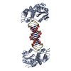
| ||||||||
| Unit cell |
|
-Components
| #1: Protein | Mass: 30023.531 Da / Num. of mol.: 1 / Fragment: N-terminal fragment (UNP residues 683-937) Source method: isolated from a genetically manipulated source Source: (gene. exp.) Moloney murine leukemia virus / Gene: gag-pro-pol / Production host:  Escherichia coli (E. coli) / References: UniProt: Q8UN00 Escherichia coli (E. coli) / References: UniProt: Q8UN00 |
|---|---|
| #2: DNA chain | Mass: 2452.647 Da / Num. of mol.: 1 / Source method: obtained synthetically / Source: (synth.) synthetic construct (others) |
| #3: DNA chain | Mass: 2536.696 Da / Num. of mol.: 1 / Source method: obtained synthetically / Source: (synth.) synthetic construct (others) |
| #4: Water | ChemComp-HOH / Water Mass: 18.015 Da / Num. of mol.: 154 / Source method: isolated from a natural source / Formula: H2O Mass: 18.015 Da / Num. of mol.: 154 / Source method: isolated from a natural source / Formula: H2O |
| Has ligand of interest | Y |
-Experimental details
-Experiment
| Experiment | Method: X-RAY DIFFRACTION / Number of used crystals: 1 |
|---|
- Sample preparation
| Crystal | Density Matthews: 2.7 Å3/Da / Density % sol: 54.49 % |
|---|---|
| Crystal grow | Temperature: 293 K / Method: vapor diffusion, hanging drop / pH: 6.5 / Details: 0.05 M ADA, pH 6.5, 9% PEG4000 |
-Data collection
| Diffraction | Mean temperature: 100 K / Serial crystal experiment: N | ||||||||||||||||||||||||||||||
|---|---|---|---|---|---|---|---|---|---|---|---|---|---|---|---|---|---|---|---|---|---|---|---|---|---|---|---|---|---|---|---|
| Diffraction source | Source: SYNCHROTRON / Site: APS / Beamline: 31-ID / Wavelength: 0.97933 Å | ||||||||||||||||||||||||||||||
| Detector | Type: DECTRIS PILATUS3 6M / Detector: PIXEL / Date: Apr 14, 2020 | ||||||||||||||||||||||||||||||
| Radiation | Protocol: SINGLE WAVELENGTH / Monochromatic (M) / Laue (L): M / Scattering type: x-ray | ||||||||||||||||||||||||||||||
| Radiation wavelength | Wavelength: 0.97933 Å / Relative weight: 1 | ||||||||||||||||||||||||||||||
| Reflection | Resolution: 1.65→145.93 Å / Num. obs: 46528 / % possible obs: 99.8 % / Redundancy: 6.5 % / Biso Wilson estimate: 28.39 Å2 / CC1/2: 0.999 / Rmerge(I) obs: 0.051 / Rpim(I) all: 0.022 / Rrim(I) all: 0.055 / Net I/σ(I): 17 / Num. measured all: 301349 / Scaling rejects: 1 | ||||||||||||||||||||||||||||||
| Reflection shell | Diffraction-ID: 1
|
- Processing
| Software |
| ||||||||||||||||||||||||||||||||||||||||||||||||||||||||||||||||||||||||||||||||||||||||||||||||||||||
|---|---|---|---|---|---|---|---|---|---|---|---|---|---|---|---|---|---|---|---|---|---|---|---|---|---|---|---|---|---|---|---|---|---|---|---|---|---|---|---|---|---|---|---|---|---|---|---|---|---|---|---|---|---|---|---|---|---|---|---|---|---|---|---|---|---|---|---|---|---|---|---|---|---|---|---|---|---|---|---|---|---|---|---|---|---|---|---|---|---|---|---|---|---|---|---|---|---|---|---|---|---|---|---|
| Refinement | Method to determine structure: MOLECULAR REPLACEMENT Starting model: PDB entry 6MIK Resolution: 1.65→27.652 Å / SU ML: 0.18 / Cross valid method: THROUGHOUT / σ(F): 1.35 / Phase error: 25.88 / Stereochemistry target values: ML
| ||||||||||||||||||||||||||||||||||||||||||||||||||||||||||||||||||||||||||||||||||||||||||||||||||||||
| Solvent computation | Shrinkage radii: 0.9 Å / VDW probe radii: 1.11 Å / Solvent model: FLAT BULK SOLVENT MODEL | ||||||||||||||||||||||||||||||||||||||||||||||||||||||||||||||||||||||||||||||||||||||||||||||||||||||
| Displacement parameters | Biso max: 105.26 Å2 / Biso mean: 41.938 Å2 / Biso min: 17.8 Å2 | ||||||||||||||||||||||||||||||||||||||||||||||||||||||||||||||||||||||||||||||||||||||||||||||||||||||
| Refinement step | Cycle: final / Resolution: 1.65→27.652 Å
| ||||||||||||||||||||||||||||||||||||||||||||||||||||||||||||||||||||||||||||||||||||||||||||||||||||||
| LS refinement shell | Refine-ID: X-RAY DIFFRACTION / Rfactor Rfree error: 0
|
