 Movie
Movie Controller


+ Open data

- Basic information
| Entry | Database: PDB / ID: 7uxv | ||||||
|---|---|---|---|---|---|---|---|
| Title | human triosephosphate isomerase mutant G122R | ||||||
 Components Components | Triosephosphate isomerase | ||||||
 Keywords Keywords | ISOMERASE / enzyme / glycolysis | ||||||
| Function / homology |  Function and homology information Function and homology informationmethylglyoxal biosynthetic process / methylglyoxal synthase / methylglyoxal synthase activity / glyceraldehyde-3-phosphate biosynthetic process / glycerol catabolic process / triose-phosphate isomerase / triose-phosphate isomerase activity / Gluconeogenesis / canonical glycolysis / Glycolysis ...methylglyoxal biosynthetic process / methylglyoxal synthase / methylglyoxal synthase activity / glyceraldehyde-3-phosphate biosynthetic process / glycerol catabolic process / triose-phosphate isomerase / triose-phosphate isomerase activity / Gluconeogenesis / canonical glycolysis / Glycolysis / gluconeogenesis / glycolytic process / ubiquitin protein ligase binding / protein homodimerization activity / extracellular space / extracellular exosome / nucleus / cytosolSimilarity search - Function | ||||||
| Biological species |  Homo sapiens (human) Homo sapiens (human) | ||||||
| Method | X-RAY DIFFRACTION / SYNCHROTRON / MOLECULAR REPLACEMENT / Resolution: 2.15 Å | ||||||
 Authors Authors | Romero, J.M. | ||||||
| Funding support |  Argentina, 1items Argentina, 1items
| ||||||
 Citation Citation | Journal: To Be Published Title: human triosephosphate isomerase mutant G122R Authors: Romero, J.M. | ||||||
| History |
|
- Structure visualization
| Structure viewer | Molecule: MolmilJmol/JSmol |
|---|
- Downloads & links
-Download
| PDBx/mmCIF format | 7uxv.cif.gz | 113.6 KB | Display | PDBx/mmCIF format |
|---|---|---|---|---|
| PDB format | pdb7uxv.ent.gz | 85.1 KB | Display | PDB format |
| PDBx/mmJSON format | 7uxv.json.gz | Tree view | PDBx/mmJSON format | |
| Others |  Other downloads Other downloads |
-Validation report
| Arichive directory | https://data.pdbj.org/pub/pdb/validation_reports/ux/7uxvftp://data.pdbj.org/pub/pdb/validation_reports/ux/7uxv | HTTPS FTP |
|---|
-Related structure data
| Related structure data | 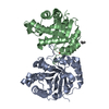 6up1S S: Starting model for refinement |
|---|---|
| Similar structure data |
-Links
 PDBj
PDBj


- Assembly
| Deposited unit | 
| ||||||||
|---|---|---|---|---|---|---|---|---|---|
| 1 |
| ||||||||
| Unit cell |
|
-Components
| #1: Protein | / TIM / Methylglyoxal synthase / Triose-phosphate isomerase Mass: 26801.607 Da / Num. of mol.: 2 / Mutation: G122R Source method: isolated from a genetically manipulated source Source: (gene. exp.) Homo sapiens (human) / Gene: TPI1, TPI / Production host:  Escherichia coli (E. coli) Escherichia coli (E. coli)References: UniProt: P60174, triose-phosphate isomerase, methylglyoxal synthase#2: Chemical |   Mass: 156.031 Da / Num. of mol.: 2 / Source method: obtained synthetically / Formula: C2H5O6P / Feature type: SUBJECT OF INVESTIGATION Mass: 156.031 Da / Num. of mol.: 2 / Source method: obtained synthetically / Formula: C2H5O6P / Feature type: SUBJECT OF INVESTIGATION#3: Chemical | ChemComp-GOL / Glycerol  Mass: 92.094 Da / Num. of mol.: 4 / Source method: obtained synthetically / Formula: C3H8O3 Mass: 92.094 Da / Num. of mol.: 4 / Source method: obtained synthetically / Formula: C3H8O3#4: Chemical | ChemComp-IPA / | Isopropyl alcohol  Mass: 60.095 Da / Num. of mol.: 1 / Source method: obtained synthetically / Formula: C3H8O / Comment: alkaloid*YM Mass: 60.095 Da / Num. of mol.: 1 / Source method: obtained synthetically / Formula: C3H8O / Comment: alkaloid*YM#5: Water | ChemComp-HOH / | Water Mass: 18.015 Da / Num. of mol.: 286 / Source method: isolated from a natural source / Formula: H2O Mass: 18.015 Da / Num. of mol.: 286 / Source method: isolated from a natural source / Formula: H2OHas ligand of interest | Y | |
|---|
-Experimental details
-Experiment
| Experiment | Method: X-RAY DIFFRACTION / Number of used crystals: 1 |
|---|
- Sample preparation
| Crystal | Density Matthews: 2.15 Å3/Da / Density % sol: 42.85 % |
|---|---|
| Crystal grow | Temperature: 283 K / Method: vapor diffusion, hanging drop / pH: 7.5 Details: 0.1 M HEPES pH 7.5, 20% PEG 4000, and 10% 2-porpanol PH range: 7.0-8.0 |
-Data collection
| Diffraction | Mean temperature: 100 K / Serial crystal experiment: N | ||||||||||||||||||||||||||||||
|---|---|---|---|---|---|---|---|---|---|---|---|---|---|---|---|---|---|---|---|---|---|---|---|---|---|---|---|---|---|---|---|
| Diffraction source | Source: SYNCHROTRON / Site: LNLS  / Beamline: W01B-MX2 / Wavelength: 1.4586 Å / Beamline: W01B-MX2 / Wavelength: 1.4586 Å | ||||||||||||||||||||||||||||||
| Detector | Type: DECTRIS PILATUS 2M / Detector: PIXEL / Date: Nov 14, 2018 | ||||||||||||||||||||||||||||||
| Radiation | Protocol: SINGLE WAVELENGTH / Monochromatic (M) / Laue (L): M / Scattering type: x-ray | ||||||||||||||||||||||||||||||
| Radiation wavelength | Wavelength: 1.4586 Å / Relative weight: 1 | ||||||||||||||||||||||||||||||
| Reflection | Resolution: 2.15→65.12 Å / Num. obs: 25450 / % possible obs: 100 % / Redundancy: 5.5 % / CC1/2: 0.996 / Rmerge(I) obs: 0.095 / Rpim(I) all: 0.044 / Rrim(I) all: 0.105 / Net I/σ(I): 11.5 / Num. measured all: 139858 / Scaling rejects: 96 | ||||||||||||||||||||||||||||||
| Reflection shell | Diffraction-ID: 1
|
- Processing
| Software |
| ||||||||||||||||||||||||||||||||||||||||||||||||||||||||||||
|---|---|---|---|---|---|---|---|---|---|---|---|---|---|---|---|---|---|---|---|---|---|---|---|---|---|---|---|---|---|---|---|---|---|---|---|---|---|---|---|---|---|---|---|---|---|---|---|---|---|---|---|---|---|---|---|---|---|---|---|---|---|
| Refinement | Method to determine structure: MOLECULAR REPLACEMENT Starting model: 6UP1 Resolution: 2.15→58.45 Å / Cor.coef. Fo:Fc: 0.95 / Cor.coef. Fo:Fc free: 0.91 / SU B: 6.418 / SU ML: 0.163 / Cross valid method: THROUGHOUT / σ(F): 0 / ESU R: 0.296 / ESU R Free: 0.221 / Stereochemistry target values: MAXIMUM LIKELIHOOD Details: HYDROGENS HAVE BEEN ADDED IN THE RIDING POSITIONS U VALUES : REFINED INDIVIDUALLY
| ||||||||||||||||||||||||||||||||||||||||||||||||||||||||||||
| Solvent computation | Ion probe radii: 0.8 Å / Shrinkage radii: 0.8 Å / VDW probe radii: 1.2 Å / Solvent model: MASK | ||||||||||||||||||||||||||||||||||||||||||||||||||||||||||||
| Displacement parameters | Biso max: 100.6 Å2 / Biso mean: 30.36 Å2 / Biso min: 13.88 Å2
| ||||||||||||||||||||||||||||||||||||||||||||||||||||||||||||
| Refinement step | Cycle: final / Resolution: 2.15→58.45 Å
| ||||||||||||||||||||||||||||||||||||||||||||||||||||||||||||
| Refine LS restraints |
| ||||||||||||||||||||||||||||||||||||||||||||||||||||||||||||
| LS refinement shell | Resolution: 2.151→2.207 Å / Rfactor Rfree error: 0 / Total num. of bins used: 20
|
