 Movie
Movie Controller


+ Open data

- Basic information
| Entry | Database: PDB / ID: 7uln | ||||||
|---|---|---|---|---|---|---|---|
| Title | Turnip yellows virus N-terminal readthrough domain | ||||||
 Components Components | Minor capsid protein P3-RTD | ||||||
 Keywords Keywords |  VIRAL PROTEIN / Viral transmission / dimer / vector interaction VIRAL PROTEIN / Viral transmission / dimer / vector interaction | ||||||
| Function / homology | Potato leaf roll virus readthrough protein / Potato leaf roll virus readthrough protein / Luteovirus group 1 coat protein / Luteovirus coat protein / host cell plasmodesma / host cell periplasmic space / viral capsid / structural molecule activity / Readthrough protein P3-RTD Function and homology information Function and homology information | ||||||
| Biological species |  Turnip yellows virus Turnip yellows virus | ||||||
| Method | X-RAY DIFFRACTION / SYNCHROTRON / MOLECULAR REPLACEMENT / molecular replacement / Resolution: 1.53 Å | ||||||
 Authors Authors | Schiltz, C.J. / Chappie, J.S. | ||||||
| Funding support |  United States, 1items United States, 1items
| ||||||
 Citation Citation | Journal: Nat Commun / Year: 2022 Title: Polerovirus N-terminal readthrough domain structures reveal molecular strategies for mitigating virus transmission by aphids Authors: Schiltz, C.J. / Wilson, J.R. / Hosford, C.J. / Adams, M.C. / Preising, S.E. / DeBlasio, S.L. / MacLeod, H.J. / Van Eck, J. / Heck, M.L. / Chappie, J.S. | ||||||
| History |
|
- Structure visualization
| Structure viewer | Molecule: MolmilJmol/JSmol |
|---|
- Downloads & links
-Download
| PDBx/mmCIF format | 7uln.cif.gz | 122 KB | Display | PDBx/mmCIF format |
|---|---|---|---|---|
| PDB format | pdb7uln.ent.gz | 92.8 KB | Display | PDB format |
| PDBx/mmJSON format | 7uln.json.gz | Tree view | PDBx/mmJSON format | |
| Others |  Other downloads Other downloads |
-Validation report
| Arichive directory | https://data.pdbj.org/pub/pdb/validation_reports/ul/7ulnftp://data.pdbj.org/pub/pdb/validation_reports/ul/7uln | HTTPS FTP |
|---|
-Related structure data
| Related structure data |  7uloSC S: Starting model for refinement C: citing same article ( |
|---|---|
| Similar structure data |
-Links
 PDBj
PDBj- Assembly
| Deposited unit | 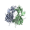
| ||||||||
|---|---|---|---|---|---|---|---|---|---|
| 1 |
| ||||||||
| Unit cell |
|
-Components
| #1: Protein | Mass: 26945.105 Da / Num. of mol.: 2 Fragment: N-terminal readthrough domain (UNP residues 224-461) Source method: isolated from a genetically manipulated source Source: (gene. exp.) Turnip yellows virus / Gene: ORF3/ORF5 / Production host:  Escherichia coli BL21(DE3) (bacteria) / References: UniProt: P09514 Escherichia coli BL21(DE3) (bacteria) / References: UniProt: P09514#2: Water | ChemComp-HOH / | Water Mass: 18.015 Da / Num. of mol.: 587 / Source method: isolated from a natural source / Formula: H2O Mass: 18.015 Da / Num. of mol.: 587 / Source method: isolated from a natural source / Formula: H2O |
|---|
-Experimental details
-Experiment
| Experiment | Method: X-RAY DIFFRACTION / Number of used crystals: 1 |
|---|
- Sample preparation
| Crystal | Density Matthews: 2.11 Å3/Da / Density % sol: 41.67 % |
|---|---|
| Crystal grow | Temperature: 293.15 K / Method: vapor diffusion, sitting drop / pH: 7.7 Details: 0.1 M HEPES, pH 7.7, 1.4 M ammonium sulfate, 0.1 M sodium chloride |
-Data collection
| Diffraction | Mean temperature: 100 K / Serial crystal experiment: N | ||||||||||||||||||||||||||||||
|---|---|---|---|---|---|---|---|---|---|---|---|---|---|---|---|---|---|---|---|---|---|---|---|---|---|---|---|---|---|---|---|
| Diffraction source | Source: SYNCHROTRON / Site: APS / Beamline: 24-ID-C / Wavelength: 0.9791 Å | ||||||||||||||||||||||||||||||
| Detector | Type: DECTRIS PILATUS 6M-F / Detector: PIXEL / Date: Apr 21, 2019 | ||||||||||||||||||||||||||||||
| Radiation | Protocol: SINGLE WAVELENGTH / Monochromatic (M) / Laue (L): M / Scattering type: x-ray | ||||||||||||||||||||||||||||||
| Radiation wavelength | Wavelength: 0.9791 Å / Relative weight: 1 | ||||||||||||||||||||||||||||||
| Reflection | Resolution: 1.53→130.78 Å / Num. obs: 69780 / % possible obs: 99.5 % / Redundancy: 6.7 % / Biso Wilson estimate: 19.33 Å2 / CC1/2: 0.999 / Rmerge(I) obs: 0.07 / Rpim(I) all: 0.029 / Rrim(I) all: 0.076 / Net I/σ(I): 20.5 | ||||||||||||||||||||||||||||||
| Reflection shell | Diffraction-ID: 1
|
-Phasing
| Phasing | Method: molecular replacement |
|---|
- Processing
| Software |
| ||||||||||||||||||||||||||||||||||||||||||||||||||||||||||||||||||||||||||||||||||||||||||||||||||||||||||||||||||||||||||||||||||||||||||||||||||||||
|---|---|---|---|---|---|---|---|---|---|---|---|---|---|---|---|---|---|---|---|---|---|---|---|---|---|---|---|---|---|---|---|---|---|---|---|---|---|---|---|---|---|---|---|---|---|---|---|---|---|---|---|---|---|---|---|---|---|---|---|---|---|---|---|---|---|---|---|---|---|---|---|---|---|---|---|---|---|---|---|---|---|---|---|---|---|---|---|---|---|---|---|---|---|---|---|---|---|---|---|---|---|---|---|---|---|---|---|---|---|---|---|---|---|---|---|---|---|---|---|---|---|---|---|---|---|---|---|---|---|---|---|---|---|---|---|---|---|---|---|---|---|---|---|---|---|---|---|---|---|---|---|
| Refinement | Method to determine structure: MOLECULAR REPLACEMENT Starting model: PDB entry 7ULO Resolution: 1.53→65.389 Å / SU ML: 0.15 / Cross valid method: THROUGHOUT / σ(F): 1.35 / Phase error: 18.85 / Stereochemistry target values: ML
| ||||||||||||||||||||||||||||||||||||||||||||||||||||||||||||||||||||||||||||||||||||||||||||||||||||||||||||||||||||||||||||||||||||||||||||||||||||||
| Solvent computation | Shrinkage radii: 0.9 Å / VDW probe radii: 1.11 Å / Solvent model: FLAT BULK SOLVENT MODEL | ||||||||||||||||||||||||||||||||||||||||||||||||||||||||||||||||||||||||||||||||||||||||||||||||||||||||||||||||||||||||||||||||||||||||||||||||||||||
| Displacement parameters | Biso max: 87.99 Å2 / Biso mean: 24.9293 Å2 / Biso min: 9.98 Å2 | ||||||||||||||||||||||||||||||||||||||||||||||||||||||||||||||||||||||||||||||||||||||||||||||||||||||||||||||||||||||||||||||||||||||||||||||||||||||
| Refinement step | Cycle: final / Resolution: 1.53→65.389 Å
| ||||||||||||||||||||||||||||||||||||||||||||||||||||||||||||||||||||||||||||||||||||||||||||||||||||||||||||||||||||||||||||||||||||||||||||||||||||||
| LS refinement shell | Refine-ID: X-RAY DIFFRACTION / Rfactor Rfree error: 0
|
