 Movie
Movie Controller

[English] 日本語
 Yorodumi
- PDB-7ugk: Crystal structure of the human queuine salvage enzyme DUF2419, wi... -
+ Open data

- Basic information
| Entry | Database: PDB / ID: 7ugk | |||||||||
|---|---|---|---|---|---|---|---|---|---|---|
| Title | Crystal structure of the human queuine salvage enzyme DUF2419, wild-type apo form | |||||||||
 Components Components | Queuosine salvage protein DUF2419 | |||||||||
 Keywords Keywords |  HYDROLASE / 7-deazaguanine salvage / queuosine / tRNA modification HYDROLASE / 7-deazaguanine salvage / queuosine / tRNA modification | |||||||||
| Function / homology | Queuosine salvage protein family / Queuosine salvage protein / tRNA-guanine transglycosylation / tRNA modification / Queuosine salvage protein Function and homology information Function and homology information | |||||||||
| Biological species |  Homo sapiens (human) Homo sapiens (human) | |||||||||
| Method | X-RAY DIFFRACTION / SYNCHROTRON / MOLECULAR REPLACEMENT / Resolution: 1.78 Å | |||||||||
 Authors Authors | Hung, S.-H. / Swairjo, M.A. | |||||||||
| Funding support |  United States, 2items United States, 2items
| |||||||||
 Citation Citation | Journal: Nucleic Acids Res. / Year: 2023 Title: Structural basis of Qng1-mediated salvage of the micronutrient queuine from queuosine-5'-monophosphate as the biological substrate. Authors: Hung, S.H. / Elliott, G.I. / Ramkumar, T.R. / Burtnyak, L. / McGrenaghan, C.J. / Alkuzweny, S. / Quaiyum, S. / Iwata-Reuyl, D. / Pan, X. / Green, B.D. / Kelly, V.P. / de Crecy-Lagard, V. / Swairjo, M.A. | |||||||||
| History |
|
- Structure visualization
| Structure viewer | Molecule: MolmilJmol/JSmol |
|---|
- Downloads & links
-Download
| PDBx/mmCIF format | 7ugk.cif.gz | 302 KB | Display | PDBx/mmCIF format |
|---|---|---|---|---|
| PDB format | pdb7ugk.ent.gz | 243.1 KB | Display | PDB format |
| PDBx/mmJSON format | 7ugk.json.gz | Tree view | PDBx/mmJSON format | |
| Others |  Other downloads Other downloads |
-Validation report
| Arichive directory | https://data.pdbj.org/pub/pdb/validation_reports/ug/7ugkftp://data.pdbj.org/pub/pdb/validation_reports/ug/7ugk | HTTPS FTP |
|---|
-Related structure data
| Related structure data |  7u07SC 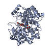 7u1oC 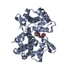 7u5aC  7u91C 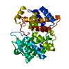 7uk3C 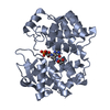 7ulcC  8dl3C S: Starting model for refinement C: citing same article ( |
|---|---|
| Similar structure data |
-Links
 PDBj
PDBj- Assembly
| Deposited unit | 
| ||||||||
|---|---|---|---|---|---|---|---|---|---|
| 1 | 
| ||||||||
| 2 | 
| ||||||||
| Unit cell |
|
-Components
| #1: Protein | Mass: 39361.777 Da / Num. of mol.: 2 Source method: isolated from a genetically manipulated source Source: (gene. exp.) Homo sapiens (human) / Gene: C9orf64 / Production host:  Escherichia coli BL21(DE3) (bacteria) / Strain (production host): BL21(DE3) / Variant (production host): C41 / References: UniProt: Q5T6V5 Escherichia coli BL21(DE3) (bacteria) / Strain (production host): BL21(DE3) / Variant (production host): C41 / References: UniProt: Q5T6V5#2: Chemical | Bis-tris methane  Mass: 209.240 Da / Num. of mol.: 2 / Source method: obtained synthetically / Formula: C8H19NO5 / Feature type: SUBJECT OF INVESTIGATION / Comment: pH buffer*YM Mass: 209.240 Da / Num. of mol.: 2 / Source method: obtained synthetically / Formula: C8H19NO5 / Feature type: SUBJECT OF INVESTIGATION / Comment: pH buffer*YM#3: Water | ChemComp-HOH / | Water Mass: 18.015 Da / Num. of mol.: 304 / Source method: isolated from a natural source / Formula: H2O Mass: 18.015 Da / Num. of mol.: 304 / Source method: isolated from a natural source / Formula: H2OHas ligand of interest | Y | |
|---|
-Experimental details
-Experiment
| Experiment | Method: X-RAY DIFFRACTION / Number of used crystals: 1 |
|---|
- Sample preparation
| Crystal | Density Matthews: 2.02 Å3/Da / Density % sol: 37.52 % / Description: Long, thin clustered crystals |
|---|---|
| Crystal grow | Temperature: 293 K / Method: vapor diffusion / pH: 5.5 / Details: 0.2M NaCl, 25% PEG3350, 0.1M Bis-Tris, pH 5.5 |
-Data collection
| Diffraction | Mean temperature: 100 K / Serial crystal experiment: N | |||||||||||||||||||||||||||||||||||||||||||||||||||||||||||||||||||||||||||||||||||||||||||||||||||||||||||||||||||||||||||||||||||||||||||||||||||||||||||||||||||||||||||||||||||||||||||||
|---|---|---|---|---|---|---|---|---|---|---|---|---|---|---|---|---|---|---|---|---|---|---|---|---|---|---|---|---|---|---|---|---|---|---|---|---|---|---|---|---|---|---|---|---|---|---|---|---|---|---|---|---|---|---|---|---|---|---|---|---|---|---|---|---|---|---|---|---|---|---|---|---|---|---|---|---|---|---|---|---|---|---|---|---|---|---|---|---|---|---|---|---|---|---|---|---|---|---|---|---|---|---|---|---|---|---|---|---|---|---|---|---|---|---|---|---|---|---|---|---|---|---|---|---|---|---|---|---|---|---|---|---|---|---|---|---|---|---|---|---|---|---|---|---|---|---|---|---|---|---|---|---|---|---|---|---|---|---|---|---|---|---|---|---|---|---|---|---|---|---|---|---|---|---|---|---|---|---|---|---|---|---|---|---|---|---|---|---|---|---|
| Diffraction source | Source: SYNCHROTRON / Site: SSRL / Beamline: BL12-2 / Wavelength: 0.97946 Å | |||||||||||||||||||||||||||||||||||||||||||||||||||||||||||||||||||||||||||||||||||||||||||||||||||||||||||||||||||||||||||||||||||||||||||||||||||||||||||||||||||||||||||||||||||||||||||||
| Detector | Type: DECTRIS PILATUS 6M / Detector: PIXEL / Date: Mar 18, 2022 | |||||||||||||||||||||||||||||||||||||||||||||||||||||||||||||||||||||||||||||||||||||||||||||||||||||||||||||||||||||||||||||||||||||||||||||||||||||||||||||||||||||||||||||||||||||||||||||
| Radiation | Protocol: SINGLE WAVELENGTH / Monochromatic (M) / Laue (L): M / Scattering type: x-ray | |||||||||||||||||||||||||||||||||||||||||||||||||||||||||||||||||||||||||||||||||||||||||||||||||||||||||||||||||||||||||||||||||||||||||||||||||||||||||||||||||||||||||||||||||||||||||||||
| Radiation wavelength | Wavelength: 0.97946 Å / Relative weight: 1 | |||||||||||||||||||||||||||||||||||||||||||||||||||||||||||||||||||||||||||||||||||||||||||||||||||||||||||||||||||||||||||||||||||||||||||||||||||||||||||||||||||||||||||||||||||||||||||||
| Reflection | Resolution: 1.78→50 Å / Num. obs: 56376 / % possible obs: 96.8 % / Redundancy: 6.6 % / Rmerge(I) obs: 0.086 / Rpim(I) all: 0.036 / Rrim(I) all: 0.094 / Χ2: 0.945 / Net I/σ(I): 9.2 / Num. measured all: 371082 | |||||||||||||||||||||||||||||||||||||||||||||||||||||||||||||||||||||||||||||||||||||||||||||||||||||||||||||||||||||||||||||||||||||||||||||||||||||||||||||||||||||||||||||||||||||||||||||
| Reflection shell | Diffraction-ID: 1
|
- Processing
| Software |
| |||||||||||||||||||||||||||||||||||||||||||||||||||||||||||||||||
|---|---|---|---|---|---|---|---|---|---|---|---|---|---|---|---|---|---|---|---|---|---|---|---|---|---|---|---|---|---|---|---|---|---|---|---|---|---|---|---|---|---|---|---|---|---|---|---|---|---|---|---|---|---|---|---|---|---|---|---|---|---|---|---|---|---|---|
| Refinement | Method to determine structure: MOLECULAR REPLACEMENT Starting model: 7U07 Resolution: 1.78→49.34 Å / Cor.coef. Fo:Fc: 0.969 / Cor.coef. Fo:Fc free: 0.961 / WRfactor Rfree: 0.1603 / WRfactor Rwork: 0.1317 / FOM work R set: 0.9393 / SU B: 3.329 / SU ML: 0.05 / SU Rfree: 0.0986 / Cross valid method: THROUGHOUT / σ(F): 0 / ESU R Free: 0.099 / Stereochemistry target values: MAXIMUM LIKELIHOOD Details: HYDROGENS HAVE BEEN ADDED IN THE RIDING POSITIONS U VALUES : REFINED INDIVIDUALLY
| |||||||||||||||||||||||||||||||||||||||||||||||||||||||||||||||||
| Solvent computation | Ion probe radii: 0.8 Å / Shrinkage radii: 0.8 Å / VDW probe radii: 1.2 Å / Solvent model: MASK | |||||||||||||||||||||||||||||||||||||||||||||||||||||||||||||||||
| Displacement parameters | Biso max: 96.5 Å2 / Biso mean: 20.888 Å2 / Biso min: 9.63 Å2
| |||||||||||||||||||||||||||||||||||||||||||||||||||||||||||||||||
| Refinement step | Cycle: final / Resolution: 1.78→49.34 Å
| |||||||||||||||||||||||||||||||||||||||||||||||||||||||||||||||||
| Refine LS restraints |
| |||||||||||||||||||||||||||||||||||||||||||||||||||||||||||||||||
| LS refinement shell | Resolution: 1.781→1.827 Å / Rfactor Rfree error: 0 / Total num. of bins used: 20
|
