 Movie
Movie Controller


+ Open data

- Basic information
| Entry | Database: PDB / ID: 7ud0 | ||||||
|---|---|---|---|---|---|---|---|
| Title | Room Temperature Drosophila Cryptochrome | ||||||
 Components Components | Cryptochrome-1 | ||||||
 Keywords Keywords | CIRCADIAN CLOCK PROTEIN / light-sensor | ||||||
| Function / homology |  Function and homology informationUV-A, blue light phototransduction / magnetoreception / detection of light stimulus involved in magnetoreception / gravitaxis / Phosphorylation of PER and TIM / Degradation of CRY / Degradation of TIM / blue light signaling pathway / response to magnetism / response to blue light ...UV-A, blue light phototransduction / magnetoreception / detection of light stimulus involved in magnetoreception / gravitaxis / Phosphorylation of PER and TIM / Degradation of CRY / Degradation of TIM / blue light signaling pathway / response to magnetism / response to blue light / regulation of circadian sleep/wake cycle, sleep / cellular response to light stimulus / blue light photoreceptor activity / entrainment of circadian clock / circadian behavior / entrainment of circadian clock by photoperiod / locomotor rhythm / photoreceptor activity / phototransduction / response to light stimulus / FAD binding / circadian regulation of gene expression / regulation of circadian rhythm / circadian rhythm / flavin adenine dinucleotide binding / negative regulation of DNA-templated transcription / perinuclear region of cytoplasm / DNA binding / nucleoplasm / nucleus / cytosol / cytoplasm Function and homology informationUV-A, blue light phototransduction / magnetoreception / detection of light stimulus involved in magnetoreception / gravitaxis / Phosphorylation of PER and TIM / Degradation of CRY / Degradation of TIM / blue light signaling pathway / response to magnetism / response to blue light ...UV-A, blue light phototransduction / magnetoreception / detection of light stimulus involved in magnetoreception / gravitaxis / Phosphorylation of PER and TIM / Degradation of CRY / Degradation of TIM / blue light signaling pathway / response to magnetism / response to blue light / regulation of circadian sleep/wake cycle, sleep / cellular response to light stimulus / blue light photoreceptor activity / entrainment of circadian clock / circadian behavior / entrainment of circadian clock by photoperiod / locomotor rhythm / photoreceptor activity / phototransduction / response to light stimulus / FAD binding / circadian regulation of gene expression / regulation of circadian rhythm / circadian rhythm / flavin adenine dinucleotide binding / negative regulation of DNA-templated transcription / perinuclear region of cytoplasm / DNA binding / nucleoplasm / nucleus / cytosol / cytoplasmSimilarity search - Function | ||||||
| Biological species |  Drosophila melanogaster (fruit fly) Drosophila melanogaster (fruit fly) | ||||||
| Method | X-RAY DIFFRACTION / SYNCHROTRON / MOLECULAR REPLACEMENT / Resolution: 3.5 Å | ||||||
 Authors Authors | Schneps, C.M. / Crane, B.R. | ||||||
| Funding support |  United States, 1items United States, 1items
| ||||||
 Citation Citation | Journal: Acta Crystallogr D Struct Biol / Year: 2022 Title: Room-temperature serial synchrotron crystallography of Drosophila cryptochrome. Authors: Schneps, C.M. / Ganguly, A. / Crane, B.R. | ||||||
| History |
|
- Structure visualization
| Structure viewer | Molecule: MolmilJmol/JSmol |
|---|
- Downloads & links
-Download
| PDBx/mmCIF format | 7ud0.cif.gz | 230.3 KB | Display | PDBx/mmCIF format |
|---|---|---|---|---|
| PDB format | pdb7ud0.ent.gz | 181.2 KB | Display | PDB format |
| PDBx/mmJSON format | 7ud0.json.gz | Tree view | PDBx/mmJSON format | |
| Others |  Other downloads Other downloads |
-Validation report
| Arichive directory | https://data.pdbj.org/pub/pdb/validation_reports/ud/7ud0ftp://data.pdbj.org/pub/pdb/validation_reports/ud/7ud0 | HTTPS FTP |
|---|
-Related structure data
| Related structure data | 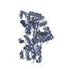 4gu5S S: Starting model for refinement |
|---|---|
| Similar structure data |
-Links
 PDBj
PDBj

- Assembly
| Deposited unit | 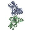
| ||||||||||||
|---|---|---|---|---|---|---|---|---|---|---|---|---|---|
| 1 | 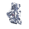
| ||||||||||||
| 2 | 
| ||||||||||||
| Unit cell |
|
-Components
| #1: Protein | / DmCRY1 / dcry / Blue light photoreceptor Mass: 62288.051 Da / Num. of mol.: 2 Source method: isolated from a genetically manipulated source Source: (gene. exp.) Drosophila melanogaster (fruit fly) / Gene: cry, CG3772 / Plasmid: pET28a(+) / Production host:  Escherichia coli (E. coli) / Strain (production host): CmpX13 / References: UniProt: O77059 Escherichia coli (E. coli) / Strain (production host): CmpX13 / References: UniProt: O77059#2: Chemical | Flavin adenine dinucleotide  Mass: 785.550 Da / Num. of mol.: 2 / Source method: obtained synthetically / Formula: C27H33N9O15P2 / Feature type: SUBJECT OF INVESTIGATION / Comment: FAD*YM Mass: 785.550 Da / Num. of mol.: 2 / Source method: obtained synthetically / Formula: C27H33N9O15P2 / Feature type: SUBJECT OF INVESTIGATION / Comment: FAD*YMHas ligand of interest | Y | |
|---|
-Experimental details
-Experiment
| Experiment | Method: X-RAY DIFFRACTION / Number of used crystals: 1 |
|---|
- Sample preparation
| Crystal | Density Matthews: 2.66 Å3/Da / Density % sol: 53.75 % |
|---|---|
| Crystal grow | Temperature: 298 K / Method: batch mode / pH: 8.5 Details: 5 mg/mL twin-strep tagged dCRY (1-539) 100 mM Tris (pH 8.5) 150 mM Magnesium acetate tetrahydrate 18% PEG-4000 Mixed 1:1 in Eppendorf tube; no vortexing |
-Data collection
| Diffraction | Mean temperature: 298 K / Ambient temp details: Room Temperature / Serial crystal experiment: Y |
|---|---|
| Diffraction source | Source: SYNCHROTRON / Site: CHESS / Beamline: A1 / Wavelength: 1.1271 Å |
| Detector | Type: DECTRIS EIGER2 S 16M / Detector: PIXEL / Date: Apr 12, 2021 |
| Radiation | Protocol: SINGLE WAVELENGTH / Monochromatic (M) / Laue (L): M / Scattering type: x-ray |
| Radiation wavelength | Wavelength: 1.1271 Å / Relative weight: 1 |
| Reflection | Resolution: 3.5→19.84 Å / Num. obs: 14881 / % possible obs: 89.8 % / Redundancy: 47.4 % / Biso Wilson estimate: 100.7 Å2 / CC1/2: 0.269 / Rmerge(I) obs: 0.649 / Rrim(I) all: 0.658 / Net I/σ(I): 5.9 |
| Reflection shell | Resolution: 3.5→3.63 Å / Redundancy: 48.6 % / Rmerge(I) obs: 2.52 / Mean I/σ(I) obs: 2 / Num. unique obs: 1462 / CC1/2: 0.213 / Rrim(I) all: 2.55 / % possible all: 89.1 |
| Serial crystallography sample delivery | Method: fixed target |
| Serial crystallography sample delivery fixed target | Description: Batch crystallization Sample dehydration prevention: Sealed chips with mylar films Sample holding: chip Sample solvent: 100 mM Tris (pH 8.5), 150 mM Magnesium acetate tetrahydrate, 18% PEG-4000 Sample unit size: 10 µm / Support base: goniometer |
- Processing
| Software |
| ||||||||||||||||||||||||
|---|---|---|---|---|---|---|---|---|---|---|---|---|---|---|---|---|---|---|---|---|---|---|---|---|---|
| Refinement | Method to determine structure: MOLECULAR REPLACEMENT Starting model: 4GU5 Resolution: 3.5→19.84 Å / Cross valid method: FREE R-VALUE Stereochemistry target values: GeoStd + Monomer Library + CDL v1.2
| ||||||||||||||||||||||||
| Displacement parameters | Biso mean: 99.65 Å2 | ||||||||||||||||||||||||
| Refinement step | Cycle: LAST / Resolution: 3.5→19.84 Å
| ||||||||||||||||||||||||
| Refine LS restraints |
| ||||||||||||||||||||||||
| LS refinement shell | Resolution: 3.5→3.63 Å
|
