 Movie
Movie Controller

[English] 日本語
 Yorodumi
- PDB-7u8k: Magic Angle Spinning NMR Structure of Human Cofilin-2 Assembled o... -
+ Open data

- Basic information
| Entry | Database: PDB / ID: 7u8k | ||||||||||||
|---|---|---|---|---|---|---|---|---|---|---|---|---|---|
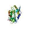
| Title | Magic Angle Spinning NMR Structure of Human Cofilin-2 Assembled on Actin Filaments | ||||||||||||
 Components Components |
| ||||||||||||
 Keywords Keywords |  CONTRACTILE PROTEIN / CELL CYCLE / actin filament binding / actin filament severing / cytoskeletal assembly CONTRACTILE PROTEIN / CELL CYCLE / actin filament binding / actin filament severing / cytoskeletal assembly | ||||||||||||
| Function / homology |  Function and homology information Function and homology informationactin filament fragmentation / positive regulation of actin filament depolymerization / actin filament severing / actin filament depolymerization / I band / sarcomere organization / cytoskeletal motor activator activity / muscle cell cellular homeostasis / tropomyosin binding / myosin heavy chain binding ...actin filament fragmentation / positive regulation of actin filament depolymerization / actin filament severing / actin filament depolymerization / I band / sarcomere organization / cytoskeletal motor activator activity / muscle cell cellular homeostasis / tropomyosin binding / myosin heavy chain binding / mesenchyme migration / troponin I binding / actin filament bundle / filamentous actin / skeletal muscle thin filament assembly / actin filament bundle assembly / striated muscle thin filament / skeletal muscle myofibril / actin monomer binding / skeletal muscle fiber development / stress fiber / skeletal muscle tissue development / titin binding / actin filament polymerization / filopodium / cell motility / actin filament / Hydrolases; Acting on acid anhydrides; Acting on acid anhydrides to facilitate cellular and subcellular movement / Z disc / nuclear matrix / calcium-dependent protein binding / actin filament binding / actin cytoskeleton / lamellipodium / cell body / hydrolase activity / protein domain specific binding / calcium ion binding / positive regulation of gene expression / magnesium ion binding / extracellular space / extracellular exosome / ATP binding / identical protein binding / cytoplasmSimilarity search - Function | ||||||||||||
| Biological species |  Oryctolagus cuniculus (rabbit) Oryctolagus cuniculus (rabbit) Homo sapiens (human) Homo sapiens (human) | ||||||||||||
| Method | SOLID-STATE NMR / molecular dynamics | ||||||||||||
 Authors Authors | Kraus, J. / Russell, R. / Kudryashova, E. / Xu, C. / Katyal, N. / Kudryashov, D. / Perilla, J.R. / Polenova, T. | ||||||||||||
| Funding support |  United States, 3items United States, 3items
| ||||||||||||
 Citation Citation | Journal: Nat Commun / Year: 2022 Title: Magic angle spinning NMR structure of human cofilin-2 assembled on actin filaments reveals isoform-specific conformation and binding mode. Authors: Kraus, J. / Russell, R.W. / Kudryashova, E. / Xu, C. / Katyal, N. / Perilla, J.R. / Kudryashov, D.S. / Polenova, T. | ||||||||||||
| History |
|
- Structure visualization
| Structure viewer | Molecule: MolmilJmol/JSmol |
|---|
- Downloads & links
-Download
| PDBx/mmCIF format | 7u8k.cif.gz | 6.1 MB | Display | PDBx/mmCIF format |
|---|---|---|---|---|
| PDB format | pdb7u8k.ent.gz | 5.1 MB | Display | PDB format |
| PDBx/mmJSON format | 7u8k.json.gz | Tree view | PDBx/mmJSON format | |
| Others |  Other downloads Other downloads |
-Validation report
| Arichive directory | https://data.pdbj.org/pub/pdb/validation_reports/u8/7u8kftp://data.pdbj.org/pub/pdb/validation_reports/u8/7u8k | HTTPS FTP |
|---|
-Related structure data
-Links
 PDBj
PDBj


- Assembly
| Deposited unit | 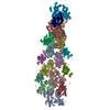
| |||||||||
|---|---|---|---|---|---|---|---|---|---|---|
| 1 |
| |||||||||
| NMR ensembles |
|
-Components
| #1: Protein | / Alpha-actin-1 Mass: 41886.660 Da / Num. of mol.: 10 Source method: isolated from a genetically manipulated source Source: (gene. exp.) Oryctolagus cuniculus (rabbit) / Gene: ACTA1, ACTA / Production host: Oryctolagus cuniculus (rabbit)References: UniProt: P68135, Hydrolases; Acting on acid anhydrides; Acting on acid anhydrides to facilitate cellular and subcellular movement#2: Protein | / Cofilin / muscle isoformMass: 18789.672 Da / Num. of mol.: 8 Source method: isolated from a genetically manipulated source Source: (gene. exp.) Homo sapiens (human) / Gene: CFL2 / Production host:  Escherichia coli (E. coli) / References: UniProt: Q9Y281 Escherichia coli (E. coli) / References: UniProt: Q9Y281 |
|---|
-Experimental details
-Experiment
| Experiment | Method: SOLID-STATE NMR | ||||||||||||||||||||||||||||||||||||||||||||||||||||||||||||||||||||||||||||||||||||
|---|---|---|---|---|---|---|---|---|---|---|---|---|---|---|---|---|---|---|---|---|---|---|---|---|---|---|---|---|---|---|---|---|---|---|---|---|---|---|---|---|---|---|---|---|---|---|---|---|---|---|---|---|---|---|---|---|---|---|---|---|---|---|---|---|---|---|---|---|---|---|---|---|---|---|---|---|---|---|---|---|---|---|---|---|---|
| NMR experiment |
|
- Sample preparation
| Details |
| ||||||||||||||||||||||||||||
|---|---|---|---|---|---|---|---|---|---|---|---|---|---|---|---|---|---|---|---|---|---|---|---|---|---|---|---|---|---|
| Sample |
| ||||||||||||||||||||||||||||
| Sample conditions | Ionic strength: 50 mM / Label: cofilin condition / pH: 6.6 / Pressure: 1 atm / Temperature: 273 K |
-NMR measurement
| NMR spectrometer | Type: Bruker AVANCE III / Manufacturer: Bruker / Model: AVANCE III / Field strength: 850 MHz |
|---|
- Processing
| NMR software |
| ||||||||||||||||||||||||||||||
|---|---|---|---|---|---|---|---|---|---|---|---|---|---|---|---|---|---|---|---|---|---|---|---|---|---|---|---|---|---|---|---|
| Refinement | Method: molecular dynamics / Software ordinal: 9 | ||||||||||||||||||||||||||||||
| NMR representative | Selection criteria: lowest energy | ||||||||||||||||||||||||||||||
| NMR ensemble | Conformer selection criteria: all calculated structures submitted Conformers calculated total number: 4 / Conformers submitted total number: 4 |
