 Movie
Movie Controller

[English] 日本語
 Yorodumi
- PDB-7twm: Structure of a borosin methyltransferase from Mycena rosella with... -
+ Open data

- Basic information
| Entry | Database: PDB / ID: 7twm | ||||||
|---|---|---|---|---|---|---|---|
| Title | Structure of a borosin methyltransferase from Mycena rosella with peptide CspL(MroMCspL) in complex with SAH | ||||||
 Components Components | MroMCspL | ||||||
 Keywords Keywords |  TRANSFERASE / RIPPs / methyltransferase / borosin / MroMA TRANSFERASE / RIPPs / methyltransferase / borosin / MroMA | ||||||
| Function / homology | S-ADENOSYL-L-HOMOCYSTEINE Function and homology information Function and homology information | ||||||
| Biological species |  Mycena rosella (fungus) Mycena rosella (fungus) | ||||||
| Method | X-RAY DIFFRACTION / SYNCHROTRON / MOLECULAR REPLACEMENT / Resolution: 1.93 Å | ||||||
 Authors Authors | Zheng, Y. / Ongpipattanakul, C. / Nair, S.K. | ||||||
| Funding support |  United States, 1items United States, 1items
| ||||||
 Citation Citation | Journal: Acs Catalysis / Year: 2022 Title: Bioconjugate Platform for Iterative Backbone N -Methylation of Peptides. Authors: Zheng, Y. / Ongpipattanakul, C. / Nair, S.K. | ||||||
| History |
|
- Structure visualization
| Structure viewer | Molecule: MolmilJmol/JSmol |
|---|
- Downloads & links
-Download
| PDBx/mmCIF format | 7twm.cif.gz | 318.5 KB | Display | PDBx/mmCIF format |
|---|---|---|---|---|
| PDB format | pdb7twm.ent.gz | 256.5 KB | Display | PDB format |
| PDBx/mmJSON format | 7twm.json.gz | Tree view | PDBx/mmJSON format | |
| Others |  Other downloads Other downloads |
-Validation report
| Arichive directory | https://data.pdbj.org/pub/pdb/validation_reports/tw/7twmftp://data.pdbj.org/pub/pdb/validation_reports/tw/7twm | HTTPS FTP |
|---|
-Related structure data
| Related structure data |  7twkSC 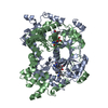 7twlC S: Starting model for refinement C: citing same article ( |
|---|---|
| Similar structure data |
-Links
 PDBj
PDBj- Assembly
| Deposited unit | 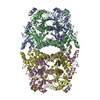
| ||||||||
|---|---|---|---|---|---|---|---|---|---|
| 1 | 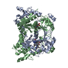
| ||||||||
| 2 | 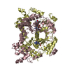
| ||||||||
| Unit cell |
|
-Components
| #1: Protein | Mass: 43996.301 Da / Num. of mol.: 4 Source method: isolated from a genetically manipulated source Source: (gene. exp.) Mycena rosella (fungus) / Production host:  Escherichia coli (E. coli) Escherichia coli (E. coli)#2: Chemical | ChemComp-SAH / S-Adenosyl-L-homocysteine  Type: L-peptide linking / Mass: 384.411 Da / Num. of mol.: 4 / Source method: obtained synthetically / Formula: C14H20N6O5S Type: L-peptide linking / Mass: 384.411 Da / Num. of mol.: 4 / Source method: obtained synthetically / Formula: C14H20N6O5S#3: Water | ChemComp-HOH / | Water Mass: 18.015 Da / Num. of mol.: 857 / Source method: isolated from a natural source / Formula: H2O Mass: 18.015 Da / Num. of mol.: 857 / Source method: isolated from a natural source / Formula: H2OHas ligand of interest | N | |
|---|
-Experimental details
-Experiment
| Experiment | Method: X-RAY DIFFRACTION / Number of used crystals: 1 |
|---|
- Sample preparation
| Crystal | Density Matthews: 2.82 Å3/Da / Density % sol: 56.33 % |
|---|---|
| Crystal grow | Temperature: 282 K / Method: vapor diffusion, hanging drop / pH: 6.5 / Details: 25% PEG 4000, and 100 mM sodium cacodylate |
-Data collection
| Diffraction | Mean temperature: 100 K / Serial crystal experiment: N |
|---|---|
| Diffraction source | Source: SYNCHROTRON / Site: APS / Beamline: 21-ID-F / Wavelength: 0.97872 Å |
| Detector | Type: RAYONIX MX-300 / Detector: CCD / Date: Jul 9, 2021 |
| Radiation | Protocol: SINGLE WAVELENGTH / Monochromatic (M) / Laue (L): M / Scattering type: x-ray |
| Radiation wavelength | Wavelength: 0.97872 Å / Relative weight: 1 |
| Reflection | Resolution: 1.93→82.7 Å / Num. obs: 145206 / % possible obs: 99.5 % / Redundancy: 13.7 % / CC1/2: 0.999 / Rmerge(I) obs: 0.109 / Net I/σ(I): 18.9 |
| Reflection shell | Resolution: 1.93→1.94 Å / Rmerge(I) obs: 1.31 / Mean I/σ(I) obs: 1.8 / Num. unique obs: 1457 / % possible all: 94 |
- Processing
| Software |
| |||||||||||||||||||||||||||||||||||||||||||||
|---|---|---|---|---|---|---|---|---|---|---|---|---|---|---|---|---|---|---|---|---|---|---|---|---|---|---|---|---|---|---|---|---|---|---|---|---|---|---|---|---|---|---|---|---|---|---|
| Refinement | Method to determine structure: MOLECULAR REPLACEMENT Starting model: 7TWK Resolution: 1.93→25 Å / Cor.coef. Fo:Fc: 0.965 / Cor.coef. Fo:Fc free: 0.953 / SU B: 3.071 / SU ML: 0.087 / Cross valid method: THROUGHOUT / σ(F): 0 / ESU R: 0.131 / ESU R Free: 0.12 / Stereochemistry target values: MAXIMUM LIKELIHOOD Details: HYDROGENS HAVE BEEN USED IF PRESENT IN THE INPUT U VALUES : REFINED INDIVIDUALLY
| |||||||||||||||||||||||||||||||||||||||||||||
| Solvent computation | Ion probe radii: 0.8 Å / Shrinkage radii: 0.8 Å / VDW probe radii: 1.2 Å / Solvent model: BABINET MODEL WITH MASK | |||||||||||||||||||||||||||||||||||||||||||||
| Displacement parameters | Biso max: 154.99 Å2 / Biso mean: 34.743 Å2 / Biso min: 15.53 Å2
| |||||||||||||||||||||||||||||||||||||||||||||
| Refinement step | Cycle: final / Resolution: 1.93→25 Å
| |||||||||||||||||||||||||||||||||||||||||||||
| Refine LS restraints |
| |||||||||||||||||||||||||||||||||||||||||||||
| LS refinement shell | Resolution: 1.932→1.982 Å / Rfactor Rfree error: 0 / Total num. of bins used: 20
|
