 Movie
Movie Controller


+ Open data

- Basic information
| Entry | Database: PDB / ID: 7tua | ||||||
|---|---|---|---|---|---|---|---|
| Title | Human Synaptotagmin-1 C2B Y312F without Ca2+ | ||||||
 Components Components | Synaptotagmin | ||||||
 Keywords Keywords | EXOCYTOSIS / C2 domain / AD3 / C2B / Greek Key / synaptotagmin | ||||||
| Function / homology |  Function and homology information Function and homology informationchromaffin granule membrane / calcium-dependent phospholipid binding / synaptic vesicle membrane / calcium ion bindingSimilarity search - Function | ||||||
| Biological species |  Homo sapiens (human) Homo sapiens (human) | ||||||
| Method | X-RAY DIFFRACTION / SYNCHROTRON / MOLECULAR REPLACEMENT / Resolution: 1.35 Å | ||||||
 Authors Authors | Dominguez, M.J. / Karmakar, S. / Fuson, K.L. / Sutton, R.B. | ||||||
| Funding support |  United States, 1items United States, 1items
| ||||||
 Citation Citation | Journal: To Be Published Title: Human Synaptotagmin-1 C2B Y312F without Ca2+ Authors: Dominguez, M.J. / Karmakar, S. / Fuson, K.L. / Sutton, R.B. #1: Journal: Acta Crystallogr., Sect. D: Biol. Crystallogr. / Year: 2012Title: Towards automated crystallographic structure refinement with phenix.refine. #2: Journal: Acta Crystallogr., Sect. D: Biol. Crystallogr. / Year: 2019Title: Macromolecular structure determination using X-rays, neutrons and electrons: recent developments in Phenix | ||||||
| History |
|
- Structure visualization
| Structure viewer | Molecule: MolmilJmol/JSmol |
|---|
- Downloads & links
-Download
| PDBx/mmCIF format | 7tua.cif.gz | 124.1 KB | Display | PDBx/mmCIF format |
|---|---|---|---|---|
| PDB format | pdb7tua.ent.gz | 87.7 KB | Display | PDB format |
| PDBx/mmJSON format | 7tua.json.gz | Tree view | PDBx/mmJSON format | |
| Others |  Other downloads Other downloads |
-Validation report
| Arichive directory | https://data.pdbj.org/pub/pdb/validation_reports/tu/7tuaftp://data.pdbj.org/pub/pdb/validation_reports/tu/7tua | HTTPS FTP |
|---|
-Related structure data
| Related structure data | 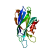 6tz3S S: Starting model for refinement |
|---|---|
| Similar structure data |
-Links
 PDBj
PDBj


- Assembly
| Deposited unit | 
| ||||||||||||
|---|---|---|---|---|---|---|---|---|---|---|---|---|---|
| 1 |
| ||||||||||||
| Unit cell |
|
-Components
| #1: Protein | Mass: 17863.818 Da / Num. of mol.: 1 / Fragment: UNP Residues 269-419 Source method: isolated from a genetically manipulated source Details: orginally cloned from human brain cDNA library / Source: (gene. exp.) Homo sapiens (human) / Tissue: brain / Gene: SYT1, hCG_2016754 / Plasmid: p202 / Production host:  Escherichia coli BL21(DE3) (bacteria) / Strain (production host): BL21(DE3) / References: UniProt: J3KQA0 Escherichia coli BL21(DE3) (bacteria) / Strain (production host): BL21(DE3) / References: UniProt: J3KQA0 |
|---|---|
| #2: Chemical | ChemComp-SO4 / Sulfate  Mass: 96.063 Da / Num. of mol.: 1 / Source method: obtained synthetically / Formula: SO4 Mass: 96.063 Da / Num. of mol.: 1 / Source method: obtained synthetically / Formula: SO4 |
| #3: Water | ChemComp-HOH / Water Mass: 18.015 Da / Num. of mol.: 153 / Source method: isolated from a natural source / Formula: H2O Mass: 18.015 Da / Num. of mol.: 153 / Source method: isolated from a natural source / Formula: H2O |
| Has ligand of interest | N |
-Experimental details
-Experiment
| Experiment | Method: X-RAY DIFFRACTION / Number of used crystals: 1 |
|---|
- Sample preparation
| Crystal | Density Matthews: 2.52 Å3/Da / Density % sol: 51.2 % |
|---|---|
| Crystal grow | Temperature: 298 K / Method: vapor diffusion, hanging drop / pH: 4.5 Details: 100 mM sodium acetate pH 4.5,200 mM ammonium sulfate, 50% w/v PEG 2000MME |
-Data collection
| Diffraction | Mean temperature: 100 K / Serial crystal experiment: N |
|---|---|
| Diffraction source | Source: SYNCHROTRON / Site: SSRL / Beamline: BL7-1 / Wavelength: 1.09717 Å |
| Detector | Type: ADSC QUANTUM 315r / Detector: CCD / Date: Feb 8, 2011 |
| Radiation | Protocol: SINGLE WAVELENGTH / Monochromatic (M) / Laue (L): M / Scattering type: x-ray |
| Radiation wavelength | Wavelength: 1.09717 Å / Relative weight: 1 |
| Reflection | Resolution: 1.35→34.69 Å / Num. obs: 39819 / % possible obs: 99.42 % / Redundancy: 13.7 % / Biso Wilson estimate: 16.57 Å2 / CC1/2: 1 / CC star: 1 / Rmerge(I) obs: 0.06954 / Rpim(I) all: 0.0176 / Rrim(I) all: 0.07183 / Net I/σ(I): 19.96 |
| Reflection shell | Resolution: 1.35→1.398 Å / Redundancy: 8.4 % / Rmerge(I) obs: 2.104 / Mean I/σ(I) obs: 1.02 / Num. unique obs: 3769 / CC1/2: 0.383 / CC star: 0.744 / Rpim(I) all: 0.7573 / Rrim(I) all: 2.243 / % possible all: 95.14 |
- Processing
| Software |
| |||||||||||||||||||||||||||||||||||||||||||||||||||||||||||||||||||||||||||||||||||||||||||||||||||||||||
|---|---|---|---|---|---|---|---|---|---|---|---|---|---|---|---|---|---|---|---|---|---|---|---|---|---|---|---|---|---|---|---|---|---|---|---|---|---|---|---|---|---|---|---|---|---|---|---|---|---|---|---|---|---|---|---|---|---|---|---|---|---|---|---|---|---|---|---|---|---|---|---|---|---|---|---|---|---|---|---|---|---|---|---|---|---|---|---|---|---|---|---|---|---|---|---|---|---|---|---|---|---|---|---|---|---|---|
| Refinement | Method to determine structure: MOLECULAR REPLACEMENT Starting model: 6tz3 Resolution: 1.35→34.69 Å / SU ML: 0.1746 / Cross valid method: FREE R-VALUE / σ(F): 1.34 / Phase error: 28.6492 Stereochemistry target values: GeoStd + Monomer Library + CDL v1.2
| |||||||||||||||||||||||||||||||||||||||||||||||||||||||||||||||||||||||||||||||||||||||||||||||||||||||||
| Solvent computation | Shrinkage radii: 0.9 Å / VDW probe radii: 1.1 Å / Solvent model: FLAT BULK SOLVENT MODEL | |||||||||||||||||||||||||||||||||||||||||||||||||||||||||||||||||||||||||||||||||||||||||||||||||||||||||
| Displacement parameters | Biso mean: 26.32 Å2 | |||||||||||||||||||||||||||||||||||||||||||||||||||||||||||||||||||||||||||||||||||||||||||||||||||||||||
| Refinement step | Cycle: LAST / Resolution: 1.35→34.69 Å
| |||||||||||||||||||||||||||||||||||||||||||||||||||||||||||||||||||||||||||||||||||||||||||||||||||||||||
| Refine LS restraints |
| |||||||||||||||||||||||||||||||||||||||||||||||||||||||||||||||||||||||||||||||||||||||||||||||||||||||||
| LS refinement shell |
|
