 Movie
Movie Controller

[English] 日本語
 Yorodumi
- PDB-7tol: X-ray crystal structure of glycerol dibiphytanyl glycerol tetraet... -
+ Open data

- Basic information
| Entry | Database: PDB / ID: 7tol | |||||||||
|---|---|---|---|---|---|---|---|---|---|---|
| Title | X-ray crystal structure of glycerol dibiphytanyl glycerol tetraether - macrocyclic archaeol synthase (GDGT-MAS) from Methanocaldococcus jannaschii with archaeal lipid, 5'deoxyadenosine, and methionine bound | |||||||||
 Components Components | glycerol dibiphytanyl glycerol tetraether - macrocyclic archaeol synthase | |||||||||
 Keywords Keywords |  OXIDOREDUCTASE / Radical SAM Enzyme / Biphytanyl Chain / Glycerol dibiphytanyl glycerol tetraether / Archaeal Lipid modification / [4Fe-4S] cluster / Rubredoxin OXIDOREDUCTASE / Radical SAM Enzyme / Biphytanyl Chain / Glycerol dibiphytanyl glycerol tetraether / Archaeal Lipid modification / [4Fe-4S] cluster / Rubredoxin | |||||||||
| Function / homology |  Function and homology informationTransferases; Transferring one-carbon groups; Methyltransferases / methyltransferase activity / 4 iron, 4 sulfur cluster binding / methylation / metal ion binding Function and homology informationTransferases; Transferring one-carbon groups; Methyltransferases / methyltransferase activity / 4 iron, 4 sulfur cluster binding / methylation / metal ion bindingSimilarity search - Function | |||||||||
| Biological species |   Methanocaldococcus jannaschii (archaea) Methanocaldococcus jannaschii (archaea) | |||||||||
| Method | X-RAY DIFFRACTION / SYNCHROTRON / MOLECULAR REPLACEMENT / Resolution: 2.03 Å | |||||||||
 Authors Authors | Lloyd, C.T. / Booker, S.J. / Boal, A.K. | |||||||||
| Funding support |  United States, 2items United States, 2items
| |||||||||
 Citation Citation | Journal: Nature / Year: 2022 Title: Discovery, structure and mechanism of a tetraether lipid synthase. Authors: Lloyd, C.T. / Iwig, D.F. / Wang, B. / Cossu, M. / Metcalf, W.W. / Boal, A.K. / Booker, S.J. | |||||||||
| History |
|
- Structure visualization
| Structure viewer | Molecule: MolmilJmol/JSmol |
|---|
- Downloads & links
-Download
| PDBx/mmCIF format | 7tol.cif.gz | 225.9 KB | Display | PDBx/mmCIF format |
|---|---|---|---|---|
| PDB format | pdb7tol.ent.gz | 175.5 KB | Display | PDB format |
| PDBx/mmJSON format | 7tol.json.gz | Tree view | PDBx/mmJSON format | |
| Others |  Other downloads Other downloads |
-Validation report
| Arichive directory | https://data.pdbj.org/pub/pdb/validation_reports/to/7tolftp://data.pdbj.org/pub/pdb/validation_reports/to/7tol | HTTPS FTP |
|---|
-Related structure data
| Related structure data |  7tomSC S: Starting model for refinement C: citing same article ( |
|---|---|
| Similar structure data |
-Links
 PDBj
PDBj
- Assembly
| Deposited unit | 
| ||||||||
|---|---|---|---|---|---|---|---|---|---|
| 1 |
| ||||||||
| Unit cell |
|
-Components
-Protein , 1 types, 1 molecules A
| #1: Protein | Mass: 59735.074 Da / Num. of mol.: 1 Source method: isolated from a genetically manipulated source Details: The first 20 residues are from a tag and are denoted with negative numbers. The 21st residue, methionine, is the start of the native protein and has sequence number 1 Source: (gene. exp.) Methanocaldococcus jannaschii (archaea)Gene: MJ0619 / Production host:  Escherichia coli BL21 (bacteria) Escherichia coli BL21 (bacteria)References: UniProt: Q58036, Transferases; Transferring one-carbon groups; Methyltransferases |
|---|
-Non-polymers , 8 types, 202 molecules 


| #2: Chemical | ChemComp-FE / Iron Mass: 55.845 Da / Num. of mol.: 1 / Source method: isolated from a natural source / Formula: Fe / Feature type: SUBJECT OF INVESTIGATION Mass: 55.845 Da / Num. of mol.: 1 / Source method: isolated from a natural source / Formula: Fe / Feature type: SUBJECT OF INVESTIGATION | ||||||||||||
|---|---|---|---|---|---|---|---|---|---|---|---|---|---|
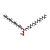
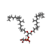
| #3: Chemical | Iron–sulfur cluster Mass: 351.640 Da / Num. of mol.: 3 / Source method: isolated from a natural source / Formula: Fe4S4 / Feature type: SUBJECT OF INVESTIGATION Mass: 351.640 Da / Num. of mol.: 3 / Source method: isolated from a natural source / Formula: Fe4S4 / Feature type: SUBJECT OF INVESTIGATION#4: Chemical | ChemComp-5AD / | Deoxyadenosine Mass: 251.242 Da / Num. of mol.: 1 / Source method: obtained synthetically / Formula: C10H13N5O3 / Feature type: SUBJECT OF INVESTIGATION Mass: 251.242 Da / Num. of mol.: 1 / Source method: obtained synthetically / Formula: C10H13N5O3 / Feature type: SUBJECT OF INVESTIGATION#5: Chemical | ChemComp-MET / | Methionine Type: L-peptide linking / Mass: 149.211 Da / Num. of mol.: 1 / Source method: obtained synthetically / Formula: C5H11NO2S / Feature type: SUBJECT OF INVESTIGATION Type: L-peptide linking / Mass: 149.211 Da / Num. of mol.: 1 / Source method: obtained synthetically / Formula: C5H11NO2S / Feature type: SUBJECT OF INVESTIGATION#6: Chemical | ChemComp-L1P / |  Mass: 733.137 Da / Num. of mol.: 1 / Source method: obtained synthetically / Formula: C43H89O6P / Feature type: SUBJECT OF INVESTIGATION Mass: 733.137 Da / Num. of mol.: 1 / Source method: obtained synthetically / Formula: C43H89O6P / Feature type: SUBJECT OF INVESTIGATION#7: Chemical | ChemComp-PGE / | Polyethylene glycol Mass: 150.173 Da / Num. of mol.: 1 / Source method: obtained synthetically / Formula: C6H14O4 Mass: 150.173 Da / Num. of mol.: 1 / Source method: obtained synthetically / Formula: C6H14O4#8: Chemical | ChemComp-L4P / |  Mass: 807.215 Da / Num. of mol.: 1 / Source method: obtained synthetically / Formula: C46H95O8P / Feature type: SUBJECT OF INVESTIGATION Mass: 807.215 Da / Num. of mol.: 1 / Source method: obtained synthetically / Formula: C46H95O8P / Feature type: SUBJECT OF INVESTIGATION#9: Water | ChemComp-HOH / | WaterMass: 18.015 Da / Num. of mol.: 193 / Source method: isolated from a natural source / Formula: H2O |
-Details
| Has ligand of interest | Y |
|---|
-Experimental details
-Experiment
| Experiment | Method: X-RAY DIFFRACTION / Number of used crystals: 1 |
|---|
- Sample preparation
| Crystal | Density Matthews: 2.25 Å3/Da / Density % sol: 45.35 % |
|---|---|
| Crystal grow | Temperature: 293 K / Method: vapor diffusion, hanging drop / pH: 6.5 / Details: MES, PEG 300, 5'deoxyadenosine and methionine |
-Data collection
| Diffraction | Mean temperature: 100 K / Serial crystal experiment: N |
|---|---|
| Diffraction source | Source: SYNCHROTRON / Site: APS / Beamline: 23-ID-B / Wavelength: 1.0332 Å |
| Detector | Type: DECTRIS EIGER X 16M / Detector: PIXEL / Date: Jul 22, 2021 |
| Radiation | Protocol: SINGLE WAVELENGTH / Monochromatic (M) / Laue (L): M / Scattering type: x-ray |
| Radiation wavelength | Wavelength: 1.0332 Å / Relative weight: 1 |
| Reflection | Resolution: 2.03→50 Å / Num. obs: 53752 / % possible obs: 98.5 % / Redundancy: 3.9 % / CC1/2: 0.987 / Net I/σ(I): 19.25 |
| Reflection shell | Resolution: 2.03→2.07 Å / Num. unique obs: 1666 / CC1/2: 0.656 |
- Processing
| Software |
| |||||||||||||||||||||||||||||||||||||||||||||||||||||||||||||||||||||||||||||||||||||||||||||||||||||||||||||||||||||||||||||||||||||||||||||||||||||||||||||||||||||||||||||||
|---|---|---|---|---|---|---|---|---|---|---|---|---|---|---|---|---|---|---|---|---|---|---|---|---|---|---|---|---|---|---|---|---|---|---|---|---|---|---|---|---|---|---|---|---|---|---|---|---|---|---|---|---|---|---|---|---|---|---|---|---|---|---|---|---|---|---|---|---|---|---|---|---|---|---|---|---|---|---|---|---|---|---|---|---|---|---|---|---|---|---|---|---|---|---|---|---|---|---|---|---|---|---|---|---|---|---|---|---|---|---|---|---|---|---|---|---|---|---|---|---|---|---|---|---|---|---|---|---|---|---|---|---|---|---|---|---|---|---|---|---|---|---|---|---|---|---|---|---|---|---|---|---|---|---|---|---|---|---|---|---|---|---|---|---|---|---|---|---|---|---|---|---|---|---|---|---|
| Refinement | Method to determine structure: MOLECULAR REPLACEMENT Starting model: 7TOM Resolution: 2.03→46.84 Å / SU ML: 0.22 / Cross valid method: THROUGHOUT / σ(F): 1.35 / Phase error: 23.32 / Stereochemistry target values: ML
| |||||||||||||||||||||||||||||||||||||||||||||||||||||||||||||||||||||||||||||||||||||||||||||||||||||||||||||||||||||||||||||||||||||||||||||||||||||||||||||||||||||||||||||||
| Solvent computation | Shrinkage radii: 0.9 Å / VDW probe radii: 1.11 Å / Solvent model: FLAT BULK SOLVENT MODEL | |||||||||||||||||||||||||||||||||||||||||||||||||||||||||||||||||||||||||||||||||||||||||||||||||||||||||||||||||||||||||||||||||||||||||||||||||||||||||||||||||||||||||||||||
| Displacement parameters | Biso max: 133.64 Å2 / Biso mean: 36.5147 Å2 / Biso min: 6.42 Å2 | |||||||||||||||||||||||||||||||||||||||||||||||||||||||||||||||||||||||||||||||||||||||||||||||||||||||||||||||||||||||||||||||||||||||||||||||||||||||||||||||||||||||||||||||
| Refinement step | Cycle: final / Resolution: 2.03→46.84 Å
| |||||||||||||||||||||||||||||||||||||||||||||||||||||||||||||||||||||||||||||||||||||||||||||||||||||||||||||||||||||||||||||||||||||||||||||||||||||||||||||||||||||||||||||||
| LS refinement shell | Refine-ID: X-RAY DIFFRACTION / Rfactor Rfree error: 0 / Total num. of bins used: 24
| |||||||||||||||||||||||||||||||||||||||||||||||||||||||||||||||||||||||||||||||||||||||||||||||||||||||||||||||||||||||||||||||||||||||||||||||||||||||||||||||||||||||||||||||
| Refinement TLS params. | Method: refined / Refine-ID: X-RAY DIFFRACTION
| |||||||||||||||||||||||||||||||||||||||||||||||||||||||||||||||||||||||||||||||||||||||||||||||||||||||||||||||||||||||||||||||||||||||||||||||||||||||||||||||||||||||||||||||
| Refinement TLS group |
|
