 Movie
Movie Controller

[English] 日本語
 Yorodumi
- PDB-7tn8: Crystal structure of Zea mays Inositol-tetrakisphosphate Kinase 1... -
+ Open data

- Basic information
| Entry | Database: PDB / ID: 7tn8 | ||||||
|---|---|---|---|---|---|---|---|
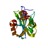
| Title | Crystal structure of Zea mays Inositol-tetrakisphosphate Kinase 1 mutant (ZmITPK1-H192A) in complex with InsP6 | ||||||
 Components Components | Inositol-tetrakisphosphate 1-kinase 1 | ||||||
 Keywords Keywords | TRANSFERASE / ATP-grasp / inositol phosphate / kinase / signal transduction | ||||||
| Function / homology |  Function and homology information Function and homology informationinositol-hexakisphosphate 5-kinase / inositol-tetrakisphosphate 1-kinase / inositol-1,3,4-trisphosphate 5/6-kinase / inositol tetrakisphosphate 1-kinase activity / inositol-1,3,4-trisphosphate 6-kinase activity / inositol-1,3,4-trisphosphate 5-kinase activity / inositol-1,3,4,5-tetrakisphosphate 3-phosphatase activity / inositol tetrakisphosphate 6-kinase activity / myo-inositol hexakisphosphate biosynthetic process / seed development ...inositol-hexakisphosphate 5-kinase / inositol-tetrakisphosphate 1-kinase / inositol-1,3,4-trisphosphate 5/6-kinase / inositol tetrakisphosphate 1-kinase activity / inositol-1,3,4-trisphosphate 6-kinase activity / inositol-1,3,4-trisphosphate 5-kinase activity / inositol-1,3,4,5-tetrakisphosphate 3-phosphatase activity / inositol tetrakisphosphate 6-kinase activity / myo-inositol hexakisphosphate biosynthetic process / seed development / inositol hexakisphosphate 5-kinase activity / inositol trisphosphate metabolic process / phosphorylation / magnesium ion binding / ATP bindingSimilarity search - Function | ||||||
| Biological species |  Zea mays (maize) Zea mays (maize) | ||||||
| Method | X-RAY DIFFRACTION / SYNCHROTRON / SAD / Resolution: 2.6 Å | ||||||
 Authors Authors | Zong, G. / Wang, H. / Shears, S.B. | ||||||
| Funding support |  United States, 1items United States, 1items
| ||||||
 Citation Citation | Journal: Faseb J. / Year: 2022 Title: Structural and catalytic analyses of the InsP 6 kinase activities of higher plant ITPKs. Authors: Zong, G. / Shears, S.B. / Wang, H. | ||||||
| History |
|
- Structure visualization
| Structure viewer | Molecule: MolmilJmol/JSmol |
|---|
- Downloads & links
-Download
| PDBx/mmCIF format | 7tn8.cif.gz | 74.4 KB | Display | PDBx/mmCIF format |
|---|---|---|---|---|
| PDB format | pdb7tn8.ent.gz | 53.1 KB | Display | PDB format |
| PDBx/mmJSON format | 7tn8.json.gz | Tree view | PDBx/mmJSON format | |
| Others |  Other downloads Other downloads |
-Validation report
| Arichive directory | https://data.pdbj.org/pub/pdb/validation_reports/tn/7tn8ftp://data.pdbj.org/pub/pdb/validation_reports/tn/7tn8 | HTTPS FTP |
|---|
-Related structure data


-Links
 PDBj
PDBj


- Assembly
| Deposited unit | 
| ||||||||
|---|---|---|---|---|---|---|---|---|---|
| 1 | 
| ||||||||
| Unit cell |
|
-Components
| #1: Protein | / Inositol 1 / 3 / 4-trisphosphate 5/6-kinase 1 / Inositol-triphosphate 5/6-kinase 1 / Ins(1 / 4)P(3) ...Inositol 1 / 3 / 4-trisphosphate 5/6-kinase 1 / Inositol-triphosphate 5/6-kinase 1 / Ins(1 / 4)P(3) 5/6-kinase 1 / Low phytic acid protein 2 / ZmIpk Mass: 37289.387 Da / Num. of mol.: 1 / Mutation: H192A Source method: isolated from a genetically manipulated source Source: (gene. exp.) Zea mays (maize) / Gene: ITPK1, LPA2 / Production host:  Escherichia coli (E. coli) Escherichia coli (E. coli)References: UniProt: Q84Y01, inositol-tetrakisphosphate 1-kinase, inositol-1,3,4-trisphosphate 5/6-kinase |
|---|---|
| #2: Chemical | ChemComp-IHP / Phytic acid  Mass: 660.035 Da / Num. of mol.: 1 / Source method: obtained synthetically / Formula: C6H18O24P6 / Feature type: SUBJECT OF INVESTIGATION Mass: 660.035 Da / Num. of mol.: 1 / Source method: obtained synthetically / Formula: C6H18O24P6 / Feature type: SUBJECT OF INVESTIGATION |
| #3: Chemical | ChemComp-CL / Chloride  Mass: 35.453 Da / Num. of mol.: 1 / Source method: obtained synthetically / Formula: Cl / Feature type: SUBJECT OF INVESTIGATION Mass: 35.453 Da / Num. of mol.: 1 / Source method: obtained synthetically / Formula: Cl / Feature type: SUBJECT OF INVESTIGATION |
| #4: Water | ChemComp-HOH / Water Mass: 18.015 Da / Num. of mol.: 95 / Source method: isolated from a natural source / Formula: H2O Mass: 18.015 Da / Num. of mol.: 95 / Source method: isolated from a natural source / Formula: H2O |
| Has ligand of interest | Y |
-Experimental details
-Experiment
| Experiment | Method: X-RAY DIFFRACTION / Number of used crystals: 1 |
|---|
- Sample preparation
| Crystal | Density Matthews: 4.09 Å3/Da / Density % sol: 69.89 % |
|---|---|
| Crystal grow | Temperature: 293 K / Method: vapor diffusion, hanging drop / pH: 7 Details: 12% PEG3350, 100 mM HEPES, 7.0, 200 mM calcium chloride, 10% glycerol PH range: 7-8 |
-Data collection
| Diffraction | Mean temperature: 100 K / Serial crystal experiment: N | |||||||||||||||||||||||||||||||||||||||||||||||||||||||||||||||||||||||||||||||||||||||||||||||||||||||||||||||||||||||||||||||||||||||||||||||||||||||||||||||||||||||||||||||||||||||||||||
|---|---|---|---|---|---|---|---|---|---|---|---|---|---|---|---|---|---|---|---|---|---|---|---|---|---|---|---|---|---|---|---|---|---|---|---|---|---|---|---|---|---|---|---|---|---|---|---|---|---|---|---|---|---|---|---|---|---|---|---|---|---|---|---|---|---|---|---|---|---|---|---|---|---|---|---|---|---|---|---|---|---|---|---|---|---|---|---|---|---|---|---|---|---|---|---|---|---|---|---|---|---|---|---|---|---|---|---|---|---|---|---|---|---|---|---|---|---|---|---|---|---|---|---|---|---|---|---|---|---|---|---|---|---|---|---|---|---|---|---|---|---|---|---|---|---|---|---|---|---|---|---|---|---|---|---|---|---|---|---|---|---|---|---|---|---|---|---|---|---|---|---|---|---|---|---|---|---|---|---|---|---|---|---|---|---|---|---|---|---|---|
| Diffraction source | Source: SYNCHROTRON / Site: APS / Beamline: 22-ID / Wavelength: 1 Å | |||||||||||||||||||||||||||||||||||||||||||||||||||||||||||||||||||||||||||||||||||||||||||||||||||||||||||||||||||||||||||||||||||||||||||||||||||||||||||||||||||||||||||||||||||||||||||||
| Detector | Type: DECTRIS EIGER X 16M / Detector: PIXEL / Date: Dec 3, 2020 | |||||||||||||||||||||||||||||||||||||||||||||||||||||||||||||||||||||||||||||||||||||||||||||||||||||||||||||||||||||||||||||||||||||||||||||||||||||||||||||||||||||||||||||||||||||||||||||
| Radiation | Protocol: SINGLE WAVELENGTH / Monochromatic (M) / Laue (L): M / Scattering type: x-ray | |||||||||||||||||||||||||||||||||||||||||||||||||||||||||||||||||||||||||||||||||||||||||||||||||||||||||||||||||||||||||||||||||||||||||||||||||||||||||||||||||||||||||||||||||||||||||||||
| Radiation wavelength | Wavelength: 1 Å / Relative weight: 1 | |||||||||||||||||||||||||||||||||||||||||||||||||||||||||||||||||||||||||||||||||||||||||||||||||||||||||||||||||||||||||||||||||||||||||||||||||||||||||||||||||||||||||||||||||||||||||||||
| Reflection | Resolution: 2.6→50 Å / Num. obs: 20094 / % possible obs: 99.7 % / Redundancy: 9.9 % / Rmerge(I) obs: 0.066 / Rpim(I) all: 0.023 / Rrim(I) all: 0.07 / Χ2: 0.97 / Net I/σ(I): 15.5 / Num. measured all: 199201 | |||||||||||||||||||||||||||||||||||||||||||||||||||||||||||||||||||||||||||||||||||||||||||||||||||||||||||||||||||||||||||||||||||||||||||||||||||||||||||||||||||||||||||||||||||||||||||||
| Reflection shell | Diffraction-ID: 1
|
- Processing
| Software |
| ||||||||||||||||||||||||||||||||||||||||||||||||||||||||||||
|---|---|---|---|---|---|---|---|---|---|---|---|---|---|---|---|---|---|---|---|---|---|---|---|---|---|---|---|---|---|---|---|---|---|---|---|---|---|---|---|---|---|---|---|---|---|---|---|---|---|---|---|---|---|---|---|---|---|---|---|---|---|
| Refinement | Method to determine structure: SAD / Resolution: 2.6→42.11 Å / Cor.coef. Fo:Fc: 0.936 / Cor.coef. Fo:Fc free: 0.911 / SU B: 7.757 / SU ML: 0.168 / Cross valid method: THROUGHOUT / σ(F): 0 / ESU R: 0.261 / ESU R Free: 0.233 / Stereochemistry target values: MAXIMUM LIKELIHOOD Details: HYDROGENS HAVE BEEN ADDED IN THE RIDING POSITIONS U VALUES : REFINED INDIVIDUALLY
| ||||||||||||||||||||||||||||||||||||||||||||||||||||||||||||
| Solvent computation | Ion probe radii: 0.8 Å / Shrinkage radii: 0.8 Å / VDW probe radii: 1.2 Å / Solvent model: MASK | ||||||||||||||||||||||||||||||||||||||||||||||||||||||||||||
| Displacement parameters | Biso max: 160.82 Å2 / Biso mean: 43.691 Å2 / Biso min: 10.08 Å2
| ||||||||||||||||||||||||||||||||||||||||||||||||||||||||||||
| Refinement step | Cycle: final / Resolution: 2.6→42.11 Å
| ||||||||||||||||||||||||||||||||||||||||||||||||||||||||||||
| Refine LS restraints |
| ||||||||||||||||||||||||||||||||||||||||||||||||||||||||||||
| LS refinement shell | Resolution: 2.6→2.663 Å / Rfactor Rfree error: 0
|
