 Movie
Movie Controller


+ Open data

- Basic information
| Entry | Database: PDB / ID: 7tmm | ||||||
|---|---|---|---|---|---|---|---|
| Title | Complete V1 Complex from Saccharomyces cerevisiae | ||||||
 Components Components |
| ||||||
 Keywords Keywords |  HYDROLASE / V-ATPase HYDROLASE / V-ATPase | ||||||
| Function / homology |  Function and homology information Function and homology informationproton-transporting V-type ATPase, V1 domain / Insulin receptor recycling / Transferrin endocytosis and recycling / ROS and RNS production in phagocytes / Amino acids regulate mTORC1 / Golgi lumen acidification / proton-transporting two-sector ATPase complex, catalytic domain / vacuolar proton-transporting V-type ATPase, V1 domain / endosomal lumen acidification / proton-transporting V-type ATPase complex ...proton-transporting V-type ATPase, V1 domain / Insulin receptor recycling / Transferrin endocytosis and recycling / ROS and RNS production in phagocytes / Amino acids regulate mTORC1 / Golgi lumen acidification / proton-transporting two-sector ATPase complex, catalytic domain / vacuolar proton-transporting V-type ATPase, V1 domain / endosomal lumen acidification / proton-transporting V-type ATPase complex / vacuolar proton-transporting V-type ATPase complex / vacuolar acidification / fungal-type vacuole membrane / vacuolar membrane / ATP metabolic process / proton-transporting ATPase activity, rotational mechanism / proton transmembrane transport / Golgi membrane / ATP bindingSimilarity search - Function | ||||||
| Biological species |  Saccharomyces cerevisiae (brewer's yeast) Saccharomyces cerevisiae (brewer's yeast) | ||||||
| Method | ELECTRON MICROSCOPY / single particle reconstruction / cryo EM / Resolution: 3.5 Å | ||||||
 Authors Authors | Vasanthakumar, T. / Keon, K.A. / Bueler, S.A. / Jaskolka, M.C. / Rubinstein, J.L. | ||||||
| Funding support |  Canada, 1items Canada, 1items
| ||||||
 Citation Citation | Journal: Nat Struct Mol Biol / Year: 2022 Title: Coordinated conformational changes in the V complex during V-ATPase reversible dissociation. Authors: Thamiya Vasanthakumar / Kristine A Keon / Stephanie A Bueler / Michael C Jaskolka / John L Rubinstein /  Abstract: Vacuolar-type ATPases (V-ATPases) are rotary enzymes that acidify intracellular compartments in eukaryotic cells. These multi-subunit complexes consist of a cytoplasmic V region that hydrolyzes ATP ...Vacuolar-type ATPases (V-ATPases) are rotary enzymes that acidify intracellular compartments in eukaryotic cells. These multi-subunit complexes consist of a cytoplasmic V region that hydrolyzes ATP and a membrane-embedded V region that transports protons. V-ATPase activity is regulated by reversible dissociation of the two regions, with the isolated V and V complexes becoming autoinhibited on disassembly and subunit C subsequently detaching from V. In yeast, assembly of the V and V regions is mediated by the regulator of the ATPase of vacuoles and endosomes (RAVE) complex through an unknown mechanism. We used cryogenic-electron microscopy of yeast V-ATPase to determine structures of the intact enzyme, the dissociated but complete V complex and the V complex lacking subunit C. On separation, V undergoes a dramatic conformational rearrangement, with its rotational state becoming incompatible for reassembly with V. Loss of subunit C allows V to match the rotational state of V, suggesting how RAVE could reassemble V and V by recruiting subunit C. | ||||||
| History |
|
- Structure visualization
| Structure viewer | Molecule: MolmilJmol/JSmol |
|---|
- Downloads & links
-Download
| PDBx/mmCIF format | 7tmm.cif.gz | 815 KB | Display | PDBx/mmCIF format |
|---|---|---|---|---|
| PDB format | pdb7tmm.ent.gz | 621.9 KB | Display | PDB format |
| PDBx/mmJSON format | 7tmm.json.gz | Tree view | PDBx/mmJSON format | |
| Others |  Other downloads Other downloads |
-Validation report
| Arichive directory | https://data.pdbj.org/pub/pdb/validation_reports/tm/7tmmftp://data.pdbj.org/pub/pdb/validation_reports/tm/7tmm | HTTPS FTP |
|---|
-Related structure data
| Related structure data |  25996MC 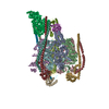 7tmoC 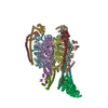 7tmpC 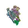 7tmqC 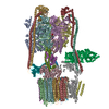 7tmrC 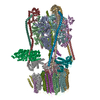 7tmsC 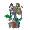 7tmtC M: map data used to model this data C: citing same article ( |
|---|---|
| Similar structure data |


-Links
 PDBj
PDBj


- Assembly
| Deposited unit | 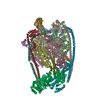
|
|---|---|
| 1 |
|
-Components
-Protein , 3 types, 9 molecules ACEBDFGIK
| #1: Protein | Mass: 70515.203 Da / Num. of mol.: 3 / Source method: isolated from a natural source / Source: (natural) Saccharomyces cerevisiae (brewer's yeast)References: UniProt: A0A6L0YX77, H+-transporting two-sector ATPase#2: Protein | Mass: 57815.023 Da / Num. of mol.: 3 / Source method: isolated from a natural source / Source: (natural) Saccharomyces cerevisiae (brewer's yeast) / References: UniProt: A0A6A5Q585#3: Protein | / HLJ1_G0040890.mRNA.1.CDS.1Mass: 26508.393 Da / Num. of mol.: 3 / Source method: isolated from a natural source / Source: (natural) Saccharomyces cerevisiae (brewer's yeast) / References: UniProt: A0A6A5Q7Y8 |
|---|
-V-type proton ATPase subunit ... , 5 types, 7 molecules HJLMNOP
| #4: Protein | Mass: 12738.706 Da / Num. of mol.: 3 / Source method: isolated from a natural source / Source: (natural) Saccharomyces cerevisiae (brewer's yeast) / References: UniProt: A0A6L0ZI53#5: Protein | | Mass: 29235.023 Da / Num. of mol.: 1 / Source method: isolated from a natural source / Source: (natural) Saccharomyces cerevisiae (brewer's yeast) / References: UniProt: A0A6A5Q1W2#6: Protein | | Mass: 13479.170 Da / Num. of mol.: 1 / Source method: isolated from a natural source / Source: (natural) Saccharomyces cerevisiae (brewer's yeast) / References: UniProt: A0A6A5PYF6#7: Protein | | Mass: 44241.352 Da / Num. of mol.: 1 / Source method: isolated from a natural source / Source: (natural) Saccharomyces cerevisiae (brewer's yeast) / References: UniProt: A0A6A5PTP1#8: Protein | | Mass: 54482.609 Da / Num. of mol.: 1 / Source method: isolated from a natural source / Source: (natural) Saccharomyces cerevisiae (brewer's yeast) / References: UniProt: P41807 |
|---|
-Non-polymers , 1 types, 1 molecules 
| #9: Chemical | ChemComp-ADP / Adenosine diphosphate Mass: 427.201 Da / Num. of mol.: 1 / Source method: obtained synthetically / Formula: C10H15N5O10P2 / Feature type: SUBJECT OF INVESTIGATION / Comment: ADP, energy-carrying molecule*YM Mass: 427.201 Da / Num. of mol.: 1 / Source method: obtained synthetically / Formula: C10H15N5O10P2 / Feature type: SUBJECT OF INVESTIGATION / Comment: ADP, energy-carrying molecule*YM |
|---|
-Details
| Has ligand of interest | Y |
|---|
-Experimental details
-Experiment
| Experiment | Method: ELECTRON MICROSCOPY |
|---|---|
| EM experiment | Aggregation state: PARTICLE / 3D reconstruction method: single particle reconstruction |
- Sample preparation
| Component | Name: Complete V1 Complex / Type: COMPLEX Details: Complete V1 complex from yeast V-ATPase following dissociation Entity ID: #1-#8 / Source: NATURAL |
|---|---|
| Molecular weight | Value: 0.635 MDa / Experimental value: NO |
| Source (natural) | Organism: Saccharomyces cerevisiae (brewer's yeast) |
| Buffer solution | pH: 7.4 |
| Specimen | Embedding applied: NO / Shadowing applied: NO / Staining applied: NO / Vitrification applied: YES |
| Vitrification | Cryogen name: ETHANE-PROPANE |
- Electron microscopy imaging
| Experimental equipment |  Model: Titan Krios / Image courtesy: FEI Company |
|---|---|
| Microscopy | Model: TFS KRIOS |
| Electron gun | Electron source: FIELD EMISSION GUN / Accelerating voltage: 300 kV / Illumination mode: FLOOD BEAM |
| Electron lens | Mode: BRIGHT FIELDBright-field microscopy / Nominal defocus max: 2000 nm / Nominal defocus min: 500 nm |
| Image recording | Electron dose: 43 e/Å2 / Film or detector model: FEI FALCON IV (4k x 4k) |
- Processing
| Software | Name: PHENIX / Version: 1.19.2_4158: / Classification: refinement | ||||||||||||||||||||||||
|---|---|---|---|---|---|---|---|---|---|---|---|---|---|---|---|---|---|---|---|---|---|---|---|---|---|
| CTF correction | Type: PHASE FLIPPING AND AMPLITUDE CORRECTION | ||||||||||||||||||||||||
| 3D reconstruction | Resolution: 3.5 Å / Resolution method: FSC 0.143 CUT-OFF / Num. of particles: 105017 / Symmetry type: POINT | ||||||||||||||||||||||||
| Refine LS restraints |
|
