 Movie
Movie Controller


+ Open data

- Basic information
| Entry | Database: PDB / ID: 7tcv | ||||||
|---|---|---|---|---|---|---|---|
| Title | VDAC K12E mutant | ||||||
 Components Components | Voltage-dependent anion-selective channel protein 1 | ||||||
 Keywords Keywords |  MEMBRANE PROTEIN / Voltage-dependent ion channel / VDAC MEMBRANE PROTEIN / Voltage-dependent ion channel / VDAC | ||||||
| Function / homology |  Function and homology informationPyruvate metabolism / negative regulation of calcium import into the mitochondrion / positive regulation of parkin-mediated stimulation of mitophagy in response to mitochondrial depolarization / voltage-gated monoatomic anion channel activity / PINK1-PRKN Mediated Mitophagy / neuron-neuron synaptic transmission / mitochondrial calcium ion transmembrane transport / acetyl-CoA biosynthetic process from pyruvate / ceramide binding / monoatomic anion channel activity ...Pyruvate metabolism / negative regulation of calcium import into the mitochondrion / positive regulation of parkin-mediated stimulation of mitophagy in response to mitochondrial depolarization / voltage-gated monoatomic anion channel activity / PINK1-PRKN Mediated Mitophagy / neuron-neuron synaptic transmission / mitochondrial calcium ion transmembrane transport / acetyl-CoA biosynthetic process from pyruvate / ceramide binding / monoatomic anion channel activity / regulation of mitophagy / mitochondrial permeability transition pore complex / phosphatidylcholine binding / Ub-specific processing proteases / oxysterol binding / cholesterol binding / porin activity / mitochondrial nucleoid / negative regulation of reactive oxygen species metabolic process / behavioral fear response / presynaptic active zone membrane / epithelial cell differentiation / learning / postsynaptic density membrane / synaptic vesicle / myelin sheath / chemical synaptic transmission / mitochondrial outer membrane / transmembrane transporter binding / mitochondrial inner membrane / membrane raft / nucleotide binding / apoptotic process / protein-containing complex binding / negative regulation of apoptotic process / protein kinase binding / protein-containing complex / mitochondrion / membrane / identical protein binding / plasma membrane Function and homology informationPyruvate metabolism / negative regulation of calcium import into the mitochondrion / positive regulation of parkin-mediated stimulation of mitophagy in response to mitochondrial depolarization / voltage-gated monoatomic anion channel activity / PINK1-PRKN Mediated Mitophagy / neuron-neuron synaptic transmission / mitochondrial calcium ion transmembrane transport / acetyl-CoA biosynthetic process from pyruvate / ceramide binding / monoatomic anion channel activity ...Pyruvate metabolism / negative regulation of calcium import into the mitochondrion / positive regulation of parkin-mediated stimulation of mitophagy in response to mitochondrial depolarization / voltage-gated monoatomic anion channel activity / PINK1-PRKN Mediated Mitophagy / neuron-neuron synaptic transmission / mitochondrial calcium ion transmembrane transport / acetyl-CoA biosynthetic process from pyruvate / ceramide binding / monoatomic anion channel activity / regulation of mitophagy / mitochondrial permeability transition pore complex / phosphatidylcholine binding / Ub-specific processing proteases / oxysterol binding / cholesterol binding / porin activity / mitochondrial nucleoid / negative regulation of reactive oxygen species metabolic process / behavioral fear response / presynaptic active zone membrane / epithelial cell differentiation / learning / postsynaptic density membrane / synaptic vesicle / myelin sheath / chemical synaptic transmission / mitochondrial outer membrane / transmembrane transporter binding / mitochondrial inner membrane / membrane raft / nucleotide binding / apoptotic process / protein-containing complex binding / negative regulation of apoptotic process / protein kinase binding / protein-containing complex / mitochondrion / membrane / identical protein binding / plasma membraneSimilarity search - Function | ||||||
| Biological species |  Mus musculus (house mouse) Mus musculus (house mouse) | ||||||
| Method | X-RAY DIFFRACTION / SYNCHROTRON / MOLECULAR REPLACEMENT / Resolution: 2.604 Å | ||||||
 Authors Authors | Khan, F. / Abramson, J. | ||||||
| Funding support | 1items
| ||||||
 Citation Citation | Journal: To Be Published Title: Dynamical control of the mitochondrial beta-barrel channel VDAC by electrostatic and mechanical coupling Authors: Ngo, V.A. / Martin, M.Q. / Khan, F. / Bergdoll, L. / Abramson, J. / Bezrukov, S.M. / Rostovtseva, T.K. / Hoogerheide, D.P. / Noskov, S.Y. | ||||||
| History |
|
- Structure visualization
| Structure viewer | Molecule: MolmilJmol/JSmol |
|---|
- Downloads & links
-Download
| PDBx/mmCIF format | 7tcv.cif.gz | 121 KB | Display | PDBx/mmCIF format |
|---|---|---|---|---|
| PDB format | pdb7tcv.ent.gz | Display | PDB format | |
| PDBx/mmJSON format | 7tcv.json.gz | Tree view | PDBx/mmJSON format | |
| Others |  Other downloads Other downloads |
-Validation report
| Arichive directory | https://data.pdbj.org/pub/pdb/validation_reports/tc/7tcvftp://data.pdbj.org/pub/pdb/validation_reports/tc/7tcv | HTTPS FTP |
|---|
-Related structure data
| Related structure data | 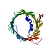 3emnS S: Starting model for refinement |
|---|---|
| Similar structure data |
-Links
 PDBj
PDBj

- Assembly
| Deposited unit | 
| ||||||||
|---|---|---|---|---|---|---|---|---|---|
| 1 | 
| ||||||||
| Unit cell |
|
-Components
| #1: Protein | Mass: 32195.879 Da / Num. of mol.: 1 / Mutation: K12E Source method: isolated from a genetically manipulated source Source: (gene. exp.) Mus musculus (house mouse) / Gene: Vdac1, Vdac5 / Production host:  Escherichia coli M15 (bacteria) / References: UniProt: Q60932 Escherichia coli M15 (bacteria) / References: UniProt: Q60932 |
|---|---|
| #2: Chemical | ChemComp-MC3 /   Mass: 677.933 Da / Num. of mol.: 1 / Source method: obtained synthetically / Formula: C36H72NO8P / Comment: phospholipid*YM Mass: 677.933 Da / Num. of mol.: 1 / Source method: obtained synthetically / Formula: C36H72NO8P / Comment: phospholipid*YM |
| #3: Water | ChemComp-HOH / Water Mass: 18.015 Da / Num. of mol.: 47 / Source method: isolated from a natural source / Formula: H2O Mass: 18.015 Da / Num. of mol.: 47 / Source method: isolated from a natural source / Formula: H2O |
| Has ligand of interest | N |
-Experimental details
-Experiment
| Experiment | Method: X-RAY DIFFRACTION / Number of used crystals: 1 |
|---|
- Sample preparation
| Crystal | Density Matthews: 3.03 Å3/Da / Density % sol: 59.47 % |
|---|---|
| Crystal grow | Temperature: 293 K / Method: vapor diffusion, hanging drop Details: 0.1 M Tris-HCl, pH 8.5, 10% Polyethylene glycol (PEG) 400 and 16% 2-Methyl-2,4-pentanediol (MPD) |
-Data collection
| Diffraction | Mean temperature: 100 K / Serial crystal experiment: N |
|---|---|
| Diffraction source | Source: SYNCHROTRON / Site: ALS  / Beamline: 5.0.1 / Wavelength: 0.9774 Å / Beamline: 5.0.1 / Wavelength: 0.9774 Å |
| Detector | Type: DECTRIS PILATUS 6M / Detector: PIXEL / Date: Apr 27, 2021 |
| Radiation | Protocol: SINGLE WAVELENGTH / Monochromatic (M) / Laue (L): M / Scattering type: x-ray |
| Radiation wavelength | Wavelength: 0.9774 Å / Relative weight: 1 |
| Reflection | Resolution: 2.6→49.745 Å / Num. obs: 11349 / % possible obs: 99 % / Redundancy: 9.8 % / CC1/2: 0.99 / Net I/σ(I): 18.7 |
| Reflection shell | Resolution: 2.6→2.72 Å / Mean I/σ(I) obs: 2.7 / Num. unique obs: 1290 / CC1/2: 0.85 |
- Processing
| Software |
| ||||||||||||||||||||||||||||||||||||||||||||||||||||||||||||||||||||||||||||||||||||||||||||||||||||||||||||||||||||||||||||||||||||||||||||||||||||||
|---|---|---|---|---|---|---|---|---|---|---|---|---|---|---|---|---|---|---|---|---|---|---|---|---|---|---|---|---|---|---|---|---|---|---|---|---|---|---|---|---|---|---|---|---|---|---|---|---|---|---|---|---|---|---|---|---|---|---|---|---|---|---|---|---|---|---|---|---|---|---|---|---|---|---|---|---|---|---|---|---|---|---|---|---|---|---|---|---|---|---|---|---|---|---|---|---|---|---|---|---|---|---|---|---|---|---|---|---|---|---|---|---|---|---|---|---|---|---|---|---|---|---|---|---|---|---|---|---|---|---|---|---|---|---|---|---|---|---|---|---|---|---|---|---|---|---|---|---|---|---|---|
| Refinement | Method to determine structure: MOLECULAR REPLACEMENT Starting model: 3EMN Resolution: 2.604→49.745 Å / Cor.coef. Fo:Fc: 0.93 / Cor.coef. Fo:Fc free: 0.892 / SU B: 10.95 / SU ML: 0.238 / Cross valid method: FREE R-VALUE / ESU R: 0.59 / ESU R Free: 0.33 Details: Hydrogens have been added in their riding positions
| ||||||||||||||||||||||||||||||||||||||||||||||||||||||||||||||||||||||||||||||||||||||||||||||||||||||||||||||||||||||||||||||||||||||||||||||||||||||
| Solvent computation | Ion probe radii: 0.8 Å / Shrinkage radii: 0.8 Å / VDW probe radii: 1.2 Å / Solvent model: MASK BULK SOLVENT | ||||||||||||||||||||||||||||||||||||||||||||||||||||||||||||||||||||||||||||||||||||||||||||||||||||||||||||||||||||||||||||||||||||||||||||||||||||||
| Displacement parameters | Biso mean: 54.627 Å2
| ||||||||||||||||||||||||||||||||||||||||||||||||||||||||||||||||||||||||||||||||||||||||||||||||||||||||||||||||||||||||||||||||||||||||||||||||||||||
| Refinement step | Cycle: LAST / Resolution: 2.604→49.745 Å
| ||||||||||||||||||||||||||||||||||||||||||||||||||||||||||||||||||||||||||||||||||||||||||||||||||||||||||||||||||||||||||||||||||||||||||||||||||||||
| Refine LS restraints |
| ||||||||||||||||||||||||||||||||||||||||||||||||||||||||||||||||||||||||||||||||||||||||||||||||||||||||||||||||||||||||||||||||||||||||||||||||||||||
| LS refinement shell |
|
