 Movie
Movie Controller

[English] 日本語
 Yorodumi
- PDB-7t6x: Cryo-EM structure of full-length hepatitis C virus E1E2 glycoprot... -
+ Open data

- Basic information
| Entry | Database: PDB / ID: 7t6x | ||||||
|---|---|---|---|---|---|---|---|
| Title | Cryo-EM structure of full-length hepatitis C virus E1E2 glycoprotein in complex with AR4A, AT12009, and IGH505 Fabs | ||||||
 Components Components |
| ||||||
 Keywords Keywords |  VIRAL PROTEIN/IMMUNE SYSTEM / hepatitis C virus / E1E2 / viral protein-immune system complex / antibody-antigen complex VIRAL PROTEIN/IMMUNE SYSTEM / hepatitis C virus / E1E2 / viral protein-immune system complex / antibody-antigen complex | ||||||
| Function / homology |  Function and homology information Function and homology informationhost cell mitochondrial membrane / host cell lipid droplet / virus-mediated perturbation of host defense response / lipid droplet / clathrin-dependent endocytosis of virus by host cell / viral nucleocapsid / host cell endoplasmic reticulum membrane / ribonucleoprotein complex / viral envelope / virion membrane ...host cell mitochondrial membrane / host cell lipid droplet / virus-mediated perturbation of host defense response / lipid droplet / clathrin-dependent endocytosis of virus by host cell / viral nucleocapsid / host cell endoplasmic reticulum membrane / ribonucleoprotein complex / viral envelope / virion membrane / structural molecule activity / membrane / cytoplasmSimilarity search - Function | ||||||
| Biological species |  Hepacivirus C Hepacivirus C Homo sapiens (human) Homo sapiens (human) | ||||||
| Method | ELECTRON MICROSCOPY / single particle reconstruction / cryo EM / Resolution: 3.83 Å | ||||||
 Authors Authors | Torrents de la Pena, A. / Ward, A.B. / Eshun-Wilson, L. / Lander, G.C. | ||||||
| Funding support |  Netherlands, 1items Netherlands, 1items
| ||||||
 Citation Citation | Journal: Science / Year: 2022 Title: Structure of the hepatitis C virus E1E2 glycoprotein complex. Authors: Alba Torrents de la Peña / Kwinten Sliepen / Lisa Eshun-Wilson / Maddy L Newby / Joel D Allen / Ian Zon / Sylvie Koekkoek / Ana Chumbe / Max Crispin / Janke Schinkel / Gabriel C Lander / ...Authors: Alba Torrents de la Peña / Kwinten Sliepen / Lisa Eshun-Wilson / Maddy L Newby / Joel D Allen / Ian Zon / Sylvie Koekkoek / Ana Chumbe / Max Crispin / Janke Schinkel / Gabriel C Lander / Rogier W Sanders / Andrew B Ward /   Abstract: Hepatitis C virus (HCV) infection is a leading cause of chronic liver disease, cirrhosis, and hepatocellular carcinoma in humans and afflicts more than 58 million people worldwide. The HCV envelope ...Hepatitis C virus (HCV) infection is a leading cause of chronic liver disease, cirrhosis, and hepatocellular carcinoma in humans and afflicts more than 58 million people worldwide. The HCV envelope E1 and E2 glycoproteins are essential for viral entry and comprise the primary antigenic target for neutralizing antibody responses. The molecular mechanisms of E1E2 assembly, as well as how the E1E2 heterodimer binds broadly neutralizing antibodies, remain elusive. Here, we present the cryo-electron microscopy structure of the membrane-extracted full-length E1E2 heterodimer in complex with three broadly neutralizing antibodies-AR4A, AT1209, and IGH505-at ~3.5-angstrom resolution. We resolve the interface between the E1 and E2 ectodomains and deliver a blueprint for the rational design of vaccine immunogens and antiviral drugs. | ||||||
| History |
|
- Structure visualization
| Structure viewer | Molecule: MolmilJmol/JSmol |
|---|
- Downloads & links
-Download
| PDBx/mmCIF format | 7t6x.cif.gz | 265.8 KB | Display | PDBx/mmCIF format |
|---|---|---|---|---|
| PDB format | pdb7t6x.ent.gz | 201.8 KB | Display | PDB format |
| PDBx/mmJSON format | 7t6x.json.gz | Tree view | PDBx/mmJSON format | |
| Others |  Other downloads Other downloads |
-Validation report
| Arichive directory | https://data.pdbj.org/pub/pdb/validation_reports/t6/7t6xftp://data.pdbj.org/pub/pdb/validation_reports/t6/7t6x | HTTPS FTP |
|---|
-Related structure data
| Related structure data | 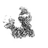 25730MC M: map data used to model this data C: citing same article ( |
|---|---|
| Similar structure data |
-Links
 PDBj
PDBj


- Assembly
| Deposited unit | 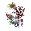
|
|---|---|
| 1 |
|
-Components
-Envelope glycoprotein ... , 2 types, 2 molecules UE
| #1: Protein | Mass: 40156.699 Da / Num. of mol.: 1 / Fragment: UNP residues 216-578 Source method: isolated from a genetically manipulated source Source: (gene. exp.) Hepacivirus C / Cell line (production host): HEK293F / Production host: Homo sapiens (human) / References: UniProt: A0A159UHX2 |
|---|---|
| #8: Protein | Mass: 20956.279 Da / Num. of mol.: 1 / Fragment: UNP residues 24-215 Source method: isolated from a genetically manipulated source Source: (gene. exp.) Hepacivirus C / Cell line (production host): HEK293F / Production host: Homo sapiens (human) / References: UniProt: A0A159UHX2 |
-Antibody , 6 types, 6 molecules HLABDC
| #2: Antibody | Mass: 29808.500 Da / Num. of mol.: 1 Source method: isolated from a genetically manipulated source Source: (gene. exp.) Homo sapiens (human) / Cell line (production host): HEK293F / Production host: Homo sapiens (human) |
|---|---|
| #3: Antibody | Mass: 25290.209 Da / Num. of mol.: 1 Source method: isolated from a genetically manipulated source Source: (gene. exp.) Homo sapiens (human) / Cell line (production host): HEK293F / Production host: Homo sapiens (human) |
| #4: Antibody | Mass: 52277.027 Da / Num. of mol.: 1 Source method: isolated from a genetically manipulated source Source: (gene. exp.) Homo sapiens (human) / Cell line (production host): HEK293F / Production host: Homo sapiens (human) |
| #5: Antibody | Mass: 24974.764 Da / Num. of mol.: 1 Source method: isolated from a genetically manipulated source Source: (gene. exp.) Homo sapiens (human) / Cell line (production host): HEK293F / Production host: Homo sapiens (human) |
| #6: Antibody | Mass: 24014.084 Da / Num. of mol.: 1 Source method: isolated from a genetically manipulated source Source: (gene. exp.) Homo sapiens (human) / Cell line (production host): HEK293F / Production host: Homo sapiens (human) |
| #7: Antibody | Mass: 29151.678 Da / Num. of mol.: 1 Source method: isolated from a genetically manipulated source Source: (gene. exp.) Homo sapiens (human) / Cell line (production host): HEK293F / Production host: Homo sapiens (human) |
-Sugars , 5 types, 15 molecules 
| #9: Polysaccharide | / Mass: 424.401 Da / Num. of mol.: 2 Source method: isolated from a genetically manipulated source #10: Polysaccharide | beta-D-mannopyranose-(1-4)-2-acetamido-2-deoxy-beta-D-glucopyranose-(1-4)-2-acetamido-2-deoxy-beta- ...beta-D-mannopyranose-(1-4)-2-acetamido-2-deoxy-beta-D-glucopyranose-(1-4)-2-acetamido-2-deoxy-beta-D-glucopyranose / Mass: 586.542 Da / Num. of mol.: 4Source method: isolated from a genetically manipulated source #11: Polysaccharide | alpha-D-mannopyranose-(1-6)-alpha-D-mannopyranose-(1-6)-beta-D-mannopyranose-(1-4)-2-acetamido-2- ...alpha-D-mannopyranose-(1-6)-alpha-D-mannopyranose-(1-6)-beta-D-mannopyranose-(1-4)-2-acetamido-2-deoxy-beta-D-glucopyranose-(1-4)-2-acetamido-2-deoxy-beta-D-glucopyranose | / Mass: 910.823 Da / Num. of mol.: 1 / Source method: obtained synthetically#12: Polysaccharide | / Mass: 910.823 Da / Num. of mol.: 2 / Source method: obtained synthetically#13: Sugar | ChemComp-NAG / N-Acetylglucosamine Type: D-saccharide, beta linking / Mass: 221.208 Da / Num. of mol.: 6 / Source method: obtained synthetically / Formula: C8H15NO6 / Feature type: SUBJECT OF INVESTIGATION Type: D-saccharide, beta linking / Mass: 221.208 Da / Num. of mol.: 6 / Source method: obtained synthetically / Formula: C8H15NO6 / Feature type: SUBJECT OF INVESTIGATION |
|---|
-Details
| Has ligand of interest | Y |
|---|
-Experimental details
-Experiment
| Experiment | Method: ELECTRON MICROSCOPY |
|---|---|
| EM experiment | Aggregation state: PARTICLE / 3D reconstruction method: single particle reconstruction |
- Sample preparation
| Component | Name: cryoEM structure of full-length hepatitis C virus E1E2 glycoprotein in complex with AR4A, AT12009 and IGH505 Fabs Type: COMPLEX / Entity ID: #1-#8 / Source: MULTIPLE SOURCES | |||||||||||||||
|---|---|---|---|---|---|---|---|---|---|---|---|---|---|---|---|---|
| Source (natural) | Organism: Hepacivirus C / Strain: AMS0232 | |||||||||||||||
| Source (recombinant) | Organism: Homo sapiens (human) / Cell: HEK 293F / Plasmid: pPPI4 | |||||||||||||||
| Buffer solution | pH: 7.4 / Details: TBS buffer, pH 7.4 | |||||||||||||||
| Buffer component |
| |||||||||||||||
| Specimen | Conc.: 0.8 mg/ml / Embedding applied: NO / Shadowing applied: NO / Staining applied: NO / Vitrification applied: YES Details: Full-length hepatitis C virus E1E2 glycoprotein complexed with AR4A, AT12009, and IGH505 Fabs and embedded in peptidiscs is diluted in TBS to a final concentration of 0.8 mg/ml. | |||||||||||||||
| Specimen support | Grid material: GOLD / Grid mesh size: 300 divisions/in. / Grid type: UltrAuFoil R1.2/1.3 | |||||||||||||||
| Vitrification | Instrument: FEI VITROBOT MARK IV / Cryogen name: ETHANE / Humidity: 100 % / Chamber temperature: 40 K Details: Blotting time: 5 s; Blotting force: 0; Waiting time: 7 s |
- Electron microscopy imaging
| Experimental equipment |  Model: Talos Arctica / Image courtesy: FEI Company |
|---|---|
| Microscopy | Model: FEI TALOS ARCTICA |
| Electron gun | Electron source: FIELD EMISSION GUN / Accelerating voltage: 200 kV / Illumination mode: FLOOD BEAM |
| Electron lens | Mode: BRIGHT FIELDBright-field microscopy / Nominal magnification: 36000 X / Calibrated magnification: 43478 X / Nominal defocus max: 1120 nm / Nominal defocus min: 800 nm / Cs: 2.7 mm / C2 aperture diameter: 70 µm / Alignment procedure: COMA FREE |
| Specimen holder | Cryogen: NITROGEN / Specimen holder model: OTHER |
| Image recording | Average exposure time: 9 sec. / Electron dose: 50 e/Å2 / Detector mode: COUNTING / Film or detector model: GATAN K2 SUMMIT (4k x 4k) / Num. of grids imaged: 2 / Num. of real images: 5121 Details: images were collected at 250 ms per frame. 36 frames were collected. |
- Processing
| Software | Name: PHENIX / Version: 1.17.1_3660: / Classification: refinement | ||||||||||||||||||||||||
|---|---|---|---|---|---|---|---|---|---|---|---|---|---|---|---|---|---|---|---|---|---|---|---|---|---|
| EM software |
| ||||||||||||||||||||||||
| CTF correction | Type: PHASE FLIPPING AND AMPLITUDE CORRECTION | ||||||||||||||||||||||||
| Particle selection | Num. of particles selected: 2017845 | ||||||||||||||||||||||||
| 3D reconstruction | Resolution: 3.83 Å / Resolution method: FSC 0.143 CUT-OFF / Num. of particles: 48546 / Symmetry type: POINT | ||||||||||||||||||||||||
| Refine LS restraints |
|
