 Movie
Movie Controller

[English] 日本語
 Yorodumi
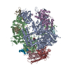
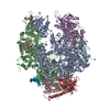
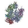
- PDB-7t2r: Structure of electron bifurcating Ni-Fe hydrogenase complex HydAB... -
+ Open data

- Basic information
| Entry | Database: PDB / ID: 7t2r | |||||||||
|---|---|---|---|---|---|---|---|---|---|---|
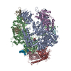
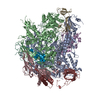
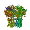
| Title | Structure of electron bifurcating Ni-Fe hydrogenase complex HydABCSL in FMN-free apo state | |||||||||
 Components Components | (NiFe hydrogenase ... ) x 5 ) x 5 | |||||||||
 Keywords Keywords | OXIDOREDUCTASE / hydrogenase complex / electron bifurcation | |||||||||
| Function / homology |  Function and homology informationferredoxin hydrogenase activity / iron-sulfur cluster binding / nickel cation binding / NADH dehydrogenase (ubiquinone) activity / ATP synthesis coupled electron transport / 2 iron, 2 sulfur cluster binding / FMN binding / 4 iron, 4 sulfur cluster binding / oxidoreductase activity / membrane / metal ion binding Function and homology informationferredoxin hydrogenase activity / iron-sulfur cluster binding / nickel cation binding / NADH dehydrogenase (ubiquinone) activity / ATP synthesis coupled electron transport / 2 iron, 2 sulfur cluster binding / FMN binding / 4 iron, 4 sulfur cluster binding / oxidoreductase activity / membrane / metal ion bindingSimilarity search - Function | |||||||||
| Biological species |  Acetomicrobium mobile (bacteria) Acetomicrobium mobile (bacteria) | |||||||||
| Method | ELECTRON MICROSCOPY / single particle reconstruction / cryo EM / Resolution: 3.2 Å | |||||||||
 Authors Authors | Feng, X. / Li, H. | |||||||||
| Funding support |  United States, 2items United States, 2items
| |||||||||
 Citation Citation | Journal: Sci Adv / Year: 2022 Title: Structure and electron transfer pathways of an electron-bifurcating NiFe-hydrogenase. Authors: Xiang Feng / Gerrit J Schut / Dominik K Haja / Michael W W Adams / Huilin Li / Abstract: Electron bifurcation enables thermodynamically unfavorable biochemical reactions. Four groups of bifurcating flavoenzyme are known and three use FAD to bifurcate. FeFe-HydABC hydrogenase represents ...Electron bifurcation enables thermodynamically unfavorable biochemical reactions. Four groups of bifurcating flavoenzyme are known and three use FAD to bifurcate. FeFe-HydABC hydrogenase represents the fourth group, but its bifurcation site is unknown. We report cryo-EM structures of the related NiFe-HydABCSL hydrogenase that reversibly oxidizes H and couples endergonic reduction of ferredoxin with exergonic reduction of NAD. FMN surrounded by a unique arrangement of iron sulfur clusters forms the bifurcating center. NAD binds to FMN in HydB, and electrons from H via HydA to a HydB [4Fe-4S] cluster enable the FMN to reduce NAD. Low-potential electron transfer from FMN to the HydC [2Fe-2S] cluster and subsequent reduction of a uniquely penta-coordinated HydB [2Fe-2S] cluster require conformational changes, leading to ferredoxin binding and reduction by a [4Fe-4S] cluster in HydB. This work clarifies the electron transfer pathways for a large group of hydrogenases underlying many essential functions in anaerobic microorganisms. | |||||||||
| History |
|
- Structure visualization
| Movie |
Movie viewer |
|---|---|
| Structure viewer | Molecule: MolmilJmol/JSmol |
- Downloads & links
-Download
| PDBx/mmCIF format | 7t2r.cif.gz | 641.1 KB | Display | PDBx/mmCIF format |
|---|---|---|---|---|
| PDB format | pdb7t2r.ent.gz | 534.2 KB | Display | PDB format |
| PDBx/mmJSON format | 7t2r.json.gz | Tree view | PDBx/mmJSON format | |
| Others |  Other downloads Other downloads |
-Validation report
| Arichive directory | https://data.pdbj.org/pub/pdb/validation_reports/t2/7t2rftp://data.pdbj.org/pub/pdb/validation_reports/t2/7t2r | HTTPS FTP |
|---|
-Related structure data
| Related structure data | 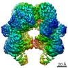 25633MC  7t30C M: map data used to model this data C: citing same article ( |
|---|---|
| Similar structure data |

-Links
 PDBj
PDBj


- Assembly
| Deposited unit | 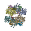
|
|---|---|
| 1 |
|
-Components
-NiFe hydrogenase ... , 5 types, 10 molecules AFBGCHDIEJ
| #1: Protein | / Anaerobic dehydrogenase Mass: 76799.648 Da / Num. of mol.: 2 / Source method: isolated from a natural source / Source: (natural) Acetomicrobium mobile (bacteria) / References: UniProt: I4BYB4#2: Protein | / NADH:ubiquinone oxidoreductase / NADH-binding (51 kD) subunitMass: 65557.148 Da / Num. of mol.: 2 / Source method: isolated from a natural source / Source: (natural) Acetomicrobium mobile (bacteria) / References: UniProt: I4BYB5#3: Protein | / NADH-quinone oxidoreductase / E subunitMass: 17482.477 Da / Num. of mol.: 2 / Source method: isolated from a natural source / Source: (natural) Acetomicrobium mobile (bacteria) / References: UniProt: I4BYB8#4: Protein | / Coenzyme F420-reducing hydrogenase / alpha subunitMass: 53294.242 Da / Num. of mol.: 2 / Source method: isolated from a natural source / Source: (natural) Acetomicrobium mobile (bacteria) / References: UniProt: I4BYB2#5: Protein | / Coenzyme F420-reducing hydrogenase / gamma subunitMass: 19965.160 Da / Num. of mol.: 2 / Source method: isolated from a natural source / Source: (natural) Acetomicrobium mobile (bacteria) / References: UniProt: I4BYB3 |
|---|
-Non-polymers , 4 types, 22 molecules 

| #6: Chemical | ChemComp-FES / Iron–sulfur cluster Mass: 175.820 Da / Num. of mol.: 6 / Source method: obtained synthetically / Formula: Fe2S2 / Feature type: SUBJECT OF INVESTIGATION Mass: 175.820 Da / Num. of mol.: 6 / Source method: obtained synthetically / Formula: Fe2S2 / Feature type: SUBJECT OF INVESTIGATION#7: Chemical | ChemComp-SF4 / Iron–sulfur cluster Mass: 351.640 Da / Num. of mol.: 12 / Source method: obtained synthetically / Formula: Fe4S4 / Feature type: SUBJECT OF INVESTIGATION Mass: 351.640 Da / Num. of mol.: 12 / Source method: obtained synthetically / Formula: Fe4S4 / Feature type: SUBJECT OF INVESTIGATION#8: Chemical | Nickel Mass: 58.693 Da / Num. of mol.: 2 / Source method: obtained synthetically / Formula: Ni / Feature type: SUBJECT OF INVESTIGATION Mass: 58.693 Da / Num. of mol.: 2 / Source method: obtained synthetically / Formula: Ni / Feature type: SUBJECT OF INVESTIGATION#9: Chemical |  Mass: 135.890 Da / Num. of mol.: 2 / Source method: obtained synthetically / Formula: C3FeN2O / Feature type: SUBJECT OF INVESTIGATION Mass: 135.890 Da / Num. of mol.: 2 / Source method: obtained synthetically / Formula: C3FeN2O / Feature type: SUBJECT OF INVESTIGATION |
|---|
-Details
| Has ligand of interest | Y |
|---|
-Experimental details
-Experiment
| Experiment | Method: ELECTRON MICROSCOPY |
|---|---|
| EM experiment | Aggregation state: PARTICLE / 3D reconstruction method: single particle reconstruction |
- Sample preparation
| Component | Name: NiFe hydrogenase complex ABCSL / Type: COMPLEX / Entity ID: #1-#5 / Source: NATURAL | |||||||||||||||
|---|---|---|---|---|---|---|---|---|---|---|---|---|---|---|---|---|
| Molecular weight | Value: 0.5 MDa / Experimental value: YES | |||||||||||||||
| Source (natural) | Organism: Acetomicrobium mobile (bacteria) | |||||||||||||||
| Buffer solution | pH: 7.5 | |||||||||||||||
| Buffer component |
| |||||||||||||||
| Specimen | Embedding applied: NO / Shadowing applied: NO / Staining applied: NO / Vitrification applied: YES | |||||||||||||||
| Specimen support | Grid material: COPPER / Grid mesh size: 300 divisions/in. / Grid type: Quantifoil R2/1 | |||||||||||||||
| Vitrification | Instrument: FEI VITROBOT MARK IV / Cryogen name: ETHANE |
- Electron microscopy imaging
| Experimental equipment |  Model: Titan Krios / Image courtesy: FEI Company |
|---|---|
| Microscopy | Model: FEI TITAN KRIOS |
| Electron gun | Electron source: FIELD EMISSION GUN / Accelerating voltage: 300 kV / Illumination mode: FLOOD BEAM |
| Electron lens | Mode: BRIGHT FIELDBright-field microscopy / Nominal defocus max: -2000 nm / Nominal defocus min: -1000 nm |
| Image recording | Electron dose: 76 e/Å2 / Detector mode: SUPER-RESOLUTION / Film or detector model: GATAN K2 SUMMIT (4k x 4k) |
- Processing
| EM software |
| ||||||||||||||||
|---|---|---|---|---|---|---|---|---|---|---|---|---|---|---|---|---|---|
| CTF correction | Type: NONE | ||||||||||||||||
| 3D reconstruction | Resolution: 3.2 Å / Resolution method: FSC 0.143 CUT-OFF / Num. of particles: 269151 / Symmetry type: POINT | ||||||||||||||||
| Atomic model building | Protocol: FLEXIBLE FIT |
