 Movie
Movie Controller

[English] 日本語
 Yorodumi
- PDB-7t1l: Crystal structure of a superbinder Fes SH2 domain (sFesS) in comp... -
+ Open data

- Basic information
| Entry | Database: PDB / ID: 7t1l | ||||||||||||
|---|---|---|---|---|---|---|---|---|---|---|---|---|---|
| Title | Crystal structure of a superbinder Fes SH2 domain (sFesS) in complex with a high affinity phosphopeptide | ||||||||||||
 Components Components |
| ||||||||||||
 Keywords Keywords |  SIGNALING PROTEIN / Src Homology 2 (SH2) / superbinder / rational engineering SIGNALING PROTEIN / Src Homology 2 (SH2) / superbinder / rational engineering | ||||||||||||
| Function / homology |  Function and homology informationterminal web assembly / intestinal D-glucose absorption / protein localization to cell cortex / establishment or maintenance of apical/basal cell polarity / regulation of microvillus length / regulation of organelle assembly / positive regulation of myeloid cell differentiation / microvillus assembly / positive regulation of early endosome to late endosome transport / membrane to membrane docking ...terminal web assembly / intestinal D-glucose absorption / protein localization to cell cortex / establishment or maintenance of apical/basal cell polarity / regulation of microvillus length / regulation of organelle assembly / positive regulation of myeloid cell differentiation / microvillus assembly / positive regulation of early endosome to late endosome transport / membrane to membrane docking / establishment of centrosome localization / negative regulation of p38MAPK cascade / Netrin-1 signaling / cortical microtubule organization / uropod / regulation of mast cell degranulation / astral microtubule organization / postsynaptic actin cytoskeleton organization / positive regulation of protein localization to early endosome / regulation of vesicle-mediated transport / SEMA3A-Plexin repulsion signaling by inhibiting Integrin adhesion / cellular response to vitamin D / positive regulation of multicellular organism growth / filopodium assembly / CRMPs in Sema3A signaling / protein-containing complex localization / microtubule bundle formation / positive regulation of monocyte differentiation / regulation of cell motility / sphingosine-1-phosphate receptor signaling pathway / establishment of endothelial barrier / S100 protein binding / Sensory processing of sound by outer hair cells of the cochlea / Sensory processing of sound by inner hair cells of the cochlea / centrosome cycle / myoblast proliferation / protein kinase A binding / cardiac muscle cell proliferation / negative regulation of interleukin-2 production / microvillus membrane / negative regulation of T cell receptor signaling pathway / leukocyte cell-cell adhesion / cortical cytoskeleton / regulation of cell differentiation / protein kinase A regulatory subunit binding / Recycling pathway of L1 / protein kinase A catalytic subunit binding / plasma membrane raft / microvillus / brush border / actin filament bundle assembly / Sema3A PAK dependent Axon repulsion / immunological synapse / regulation of cell adhesion / immunoglobulin receptor binding / cellular response to cAMP / positive regulation of microtubule polymerization / cell adhesion molecule binding / ruffle / protein kinase A signaling / phosphatidylinositol binding / ciliary basal body / filopodium / cell projection / protein localization to plasma membrane / cell periphery / actin filament / positive regulation of protein localization to plasma membrane / adherens junction / non-specific protein-tyrosine kinase / non-membrane spanning protein tyrosine kinase activity / Signaling by SCF-KIT / receptor internalization / negative regulation of ERK1 and ERK2 cascade / cytoplasmic side of plasma membrane / fibrillar center / ruffle membrane / positive regulation of neuron projection development / peptidyl-tyrosine phosphorylation / positive regulation of protein catabolic process / chemotaxis / microtubule cytoskeleton / disordered domain specific binding / actin filament binding / actin cytoskeleton / apical part of cell / actin binding / regulation of cell population proliferation / regulation of cell shape / ATPase binding / cytoplasmic vesicle / actin cytoskeleton organization / microtubule binding / basolateral plasma membrane / protein tyrosine kinase activity / vesicle / protein autophosphorylation / cell adhesion / endosome / cadherin binding Function and homology informationterminal web assembly / intestinal D-glucose absorption / protein localization to cell cortex / establishment or maintenance of apical/basal cell polarity / regulation of microvillus length / regulation of organelle assembly / positive regulation of myeloid cell differentiation / microvillus assembly / positive regulation of early endosome to late endosome transport / membrane to membrane docking ...terminal web assembly / intestinal D-glucose absorption / protein localization to cell cortex / establishment or maintenance of apical/basal cell polarity / regulation of microvillus length / regulation of organelle assembly / positive regulation of myeloid cell differentiation / microvillus assembly / positive regulation of early endosome to late endosome transport / membrane to membrane docking / establishment of centrosome localization / negative regulation of p38MAPK cascade / Netrin-1 signaling / cortical microtubule organization / uropod / regulation of mast cell degranulation / astral microtubule organization / postsynaptic actin cytoskeleton organization / positive regulation of protein localization to early endosome / regulation of vesicle-mediated transport / SEMA3A-Plexin repulsion signaling by inhibiting Integrin adhesion / cellular response to vitamin D / positive regulation of multicellular organism growth / filopodium assembly / CRMPs in Sema3A signaling / protein-containing complex localization / microtubule bundle formation / positive regulation of monocyte differentiation / regulation of cell motility / sphingosine-1-phosphate receptor signaling pathway / establishment of endothelial barrier / S100 protein binding / Sensory processing of sound by outer hair cells of the cochlea / Sensory processing of sound by inner hair cells of the cochlea / centrosome cycle / myoblast proliferation / protein kinase A binding / cardiac muscle cell proliferation / negative regulation of interleukin-2 production / microvillus membrane / negative regulation of T cell receptor signaling pathway / leukocyte cell-cell adhesion / cortical cytoskeleton / regulation of cell differentiation / protein kinase A regulatory subunit binding / Recycling pathway of L1 / protein kinase A catalytic subunit binding / plasma membrane raft / microvillus / brush border / actin filament bundle assembly / Sema3A PAK dependent Axon repulsion / immunological synapse / regulation of cell adhesion / immunoglobulin receptor binding / cellular response to cAMP / positive regulation of microtubule polymerization / cell adhesion molecule binding / ruffle / protein kinase A signaling / phosphatidylinositol binding / ciliary basal body / filopodium / cell projection / protein localization to plasma membrane / cell periphery / actin filament / positive regulation of protein localization to plasma membrane / adherens junction / non-specific protein-tyrosine kinase / non-membrane spanning protein tyrosine kinase activity / Signaling by SCF-KIT / receptor internalization / negative regulation of ERK1 and ERK2 cascade / cytoplasmic side of plasma membrane / fibrillar center / ruffle membrane / positive regulation of neuron projection development / peptidyl-tyrosine phosphorylation / positive regulation of protein catabolic process / chemotaxis / microtubule cytoskeleton / disordered domain specific binding / actin filament binding / actin cytoskeleton / apical part of cell / actin binding / regulation of cell population proliferation / regulation of cell shape / ATPase binding / cytoplasmic vesicle / actin cytoskeleton organization / microtubule binding / basolateral plasma membrane / protein tyrosine kinase activity / vesicle / protein autophosphorylation / cell adhesion / endosome / cadherin bindingSimilarity search - Function | ||||||||||||
| Biological species |  Homo sapiens (human) Homo sapiens (human) | ||||||||||||
| Method | X-RAY DIFFRACTION / SYNCHROTRON / MOLECULAR REPLACEMENT / Resolution: 1.35 Å | ||||||||||||
 Authors Authors | Martyn, G.D. / Singer, A.U. / Veggiani, G. / Kurinov, I. / Sicheri, F. / Sidhu, S.S. | ||||||||||||
| Funding support |  United States, 3items United States, 3items
| ||||||||||||
 Citation Citation | Journal: Acs Chem.Biol. / Year: 2022 Title: Engineered SH2 Domains for Targeted Phosphoproteomics. Authors: Martyn, G.D. / Veggiani, G. / Kusebauch, U. / Morrone, S.R. / Yates, B.P. / Singer, A.U. / Tong, J. / Manczyk, N. / Gish, G. / Sun, Z. / Kurinov, I. / Sicheri, F. / Moran, M.F. / Moritz, R.L. / Sidhu, S.S. | ||||||||||||
| History |
|
- Structure visualization
| Structure viewer | Molecule: MolmilJmol/JSmol |
|---|
- Downloads & links
-Download
| PDBx/mmCIF format | 7t1l.cif.gz | 157.1 KB | Display | PDBx/mmCIF format |
|---|---|---|---|---|
| PDB format | pdb7t1l.ent.gz | 111.4 KB | Display | PDB format |
| PDBx/mmJSON format | 7t1l.json.gz | Tree view | PDBx/mmJSON format | |
| Others |  Other downloads Other downloads |
-Validation report
| Arichive directory | https://data.pdbj.org/pub/pdb/validation_reports/t1/7t1lftp://data.pdbj.org/pub/pdb/validation_reports/t1/7t1l | HTTPS FTP |
|---|
-Related structure data
| Related structure data |  7t1kC  7t1uC  3bkbS S: Starting model for refinement C: citing same article ( |
|---|---|
| Similar structure data |
-Links
 PDBj
PDBj


- Assembly
| Deposited unit | 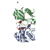
| ||||||||||||
|---|---|---|---|---|---|---|---|---|---|---|---|---|---|
| 1 | 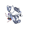
| ||||||||||||
| 2 | 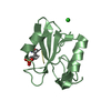
| ||||||||||||
| Unit cell |
|
-Components
| #1: Protein | Mass: 11201.869 Da / Num. of mol.: 2 / Fragment: SH2 domain Mutation: Residues 486 to 492 mutated from QGKQEYV to ETVKGAYA, and I505L (Uniprot numbering) Source method: isolated from a genetically manipulated source Source: (gene. exp.) Homo sapiens (human) / Gene: FES, FPS / Plasmid: pHH1028 / Details (production host): Nterm: His6x-TEVcleavage / Production host:  Escherichia coli BL21(DE3) (bacteria) Escherichia coli BL21(DE3) (bacteria)References: UniProt: P07332, non-specific protein-tyrosine kinase#2: Protein/peptide | Mass: 879.888 Da / Num. of mol.: 2 / Source method: obtained synthetically / Source: (synth.) Homo sapiens (human) / References: UniProt: P15311#3: Chemical | ChemComp-NA / |   Mass: 22.990 Da / Num. of mol.: 1 / Source method: obtained synthetically / Formula: Na Mass: 22.990 Da / Num. of mol.: 1 / Source method: obtained synthetically / Formula: Na#4: Chemical | ChemComp-CL / | Chloride  Mass: 35.453 Da / Num. of mol.: 1 / Source method: obtained synthetically / Formula: Cl Mass: 35.453 Da / Num. of mol.: 1 / Source method: obtained synthetically / Formula: Cl#5: Water | ChemComp-HOH / | Water Mass: 18.015 Da / Num. of mol.: 181 / Source method: isolated from a natural source / Formula: H2O Mass: 18.015 Da / Num. of mol.: 181 / Source method: isolated from a natural source / Formula: H2OHas ligand of interest | Y | |
|---|
-Experimental details
-Experiment
| Experiment | Method: X-RAY DIFFRACTION / Number of used crystals: 1 |
|---|
- Sample preparation
| Crystal | Density Matthews: 1.87 Å3/Da / Density % sol: 34.35 % |
|---|---|
| Crystal grow | Temperature: 293 K / Method: vapor diffusion, sitting drop / pH: 8.5 Details: 1.3 M Sodium Citrate Tribasic Dihydrate and 100 mM Bis-Tris Propane (pH=8.5) Temp details: Room temperature |
-Data collection
| Diffraction | Mean temperature: 100 K / Serial crystal experiment: N |
|---|---|
| Diffraction source | Source: SYNCHROTRON / Site: APS / Beamline: 24-ID-E / Wavelength: 0.97918 Å |
| Detector | Type: DECTRIS EIGER X 16M / Detector: PIXEL / Date: Jul 19, 2019 / Details: mirrors |
| Radiation | Monochromator: Si(220) / Protocol: SINGLE WAVELENGTH / Monochromatic (M) / Laue (L): M / Scattering type: x-ray |
| Radiation wavelength | Wavelength: 0.97918 Å / Relative weight: 1 |
| Reflection | Resolution: 1.3→43.32 Å / Num. obs: 45098 / % possible obs: 99.4 % / Redundancy: 6.4 % / Biso Wilson estimate: 17.86 Å2 / CC1/2: 0.998 / Rmerge(I) obs: 0.062 / Net I/σ(I): 12 |
| Reflection shell | Resolution: 1.3→1.32 Å / Redundancy: 6.6 % / Rmerge(I) obs: 0.758 / Num. unique obs: 2269 / CC1/2: 0.723 / % possible all: 98.6 |
- Processing
| Software |
| ||||||||||||||||||||||||||||||||||||||||||||||||||||||||||||||||||||||||||||||||||||||||||||||||||
|---|---|---|---|---|---|---|---|---|---|---|---|---|---|---|---|---|---|---|---|---|---|---|---|---|---|---|---|---|---|---|---|---|---|---|---|---|---|---|---|---|---|---|---|---|---|---|---|---|---|---|---|---|---|---|---|---|---|---|---|---|---|---|---|---|---|---|---|---|---|---|---|---|---|---|---|---|---|---|---|---|---|---|---|---|---|---|---|---|---|---|---|---|---|---|---|---|---|---|---|
| Refinement | Method to determine structure: MOLECULAR REPLACEMENT Starting model: 3BKB Resolution: 1.35→43.32 Å / SU ML: 0.1474 / Cross valid method: FREE R-VALUE / σ(F): 1.35 / Phase error: 19.7741 Stereochemistry target values: GeoStd + Monomer Library + CDL v1.2
| ||||||||||||||||||||||||||||||||||||||||||||||||||||||||||||||||||||||||||||||||||||||||||||||||||
| Solvent computation | Shrinkage radii: 0.9 Å / VDW probe radii: 1.11 Å / Solvent model: FLAT BULK SOLVENT MODEL | ||||||||||||||||||||||||||||||||||||||||||||||||||||||||||||||||||||||||||||||||||||||||||||||||||
| Displacement parameters | Biso mean: 22.59 Å2 | ||||||||||||||||||||||||||||||||||||||||||||||||||||||||||||||||||||||||||||||||||||||||||||||||||
| Refinement step | Cycle: LAST / Resolution: 1.35→43.32 Å
| ||||||||||||||||||||||||||||||||||||||||||||||||||||||||||||||||||||||||||||||||||||||||||||||||||
| Refine LS restraints |
| ||||||||||||||||||||||||||||||||||||||||||||||||||||||||||||||||||||||||||||||||||||||||||||||||||
| LS refinement shell |
|
