 Movie
Movie Controller


+ Open data

- Basic information
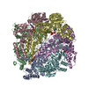
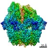
| Entry | Database: PDB / ID: 7sxo | ||||||
|---|---|---|---|---|---|---|---|
| Title | Yeast Lon (PIM1) with endogenous substrate | ||||||
 Components Components |
| ||||||
 Keywords Keywords |  HYDROLASE / Protease / AAA+ ATPase HYDROLASE / Protease / AAA+ ATPase | ||||||
| Function / homology |  Function and homology information Function and homology informationregulation of mitochondrial DNA metabolic process / oxidation-dependent protein catabolic process / mitochondrial respiratory chain complex assembly / endopeptidase La / mitochondrial DNA metabolic process / ATP-dependent peptidase activity / protein quality control for misfolded or incompletely synthesized proteins / chaperone-mediated protein complex assembly / mitochondrion organization / protein folding ...regulation of mitochondrial DNA metabolic process / oxidation-dependent protein catabolic process / mitochondrial respiratory chain complex assembly / endopeptidase La / mitochondrial DNA metabolic process / ATP-dependent peptidase activity / protein quality control for misfolded or incompletely synthesized proteins / chaperone-mediated protein complex assembly / mitochondrion organization / protein folding / single-stranded DNA binding / cellular response to oxidative stress / response to heat / sequence-specific DNA binding / mitochondrial matrix / serine-type endopeptidase activity / ATP hydrolysis activity / mitochondrion / ATP bindingSimilarity search - Function | ||||||
| Biological species |  Saccharomyces cerevisiae (brewer's yeast) Saccharomyces cerevisiae (brewer's yeast) Escherichia coli (E. coli) Escherichia coli (E. coli) | ||||||
| Method | ELECTRON MICROSCOPY / single particle reconstruction / cryo EM / Resolution: 3.3 Å | ||||||
 Authors Authors | Yang, J. / Song, A.S. / Wiseman, R.L. / Lander, G.C. | ||||||
| Funding support |  United States, 1items United States, 1items
| ||||||
 Citation Citation | Journal: J Biol Chem / Year: 2022 Title: Cryo-EM structure of hexameric yeast Lon protease (PIM1) highlights the importance of conserved structural elements. Authors: Jie Yang / Albert S Song / R Luke Wiseman / Gabriel C Lander / Abstract: Lon protease is a conserved ATP-dependent serine protease composed of an AAA+ domain that mechanically unfolds substrates and a serine protease domain that degrades these unfolded substrates. In ...Lon protease is a conserved ATP-dependent serine protease composed of an AAA+ domain that mechanically unfolds substrates and a serine protease domain that degrades these unfolded substrates. In yeast, dysregulation of Lon protease (PIM1) attenuates lifespan and leads to gross mitochondrial morphological perturbations. Although structures of the bacterial and human Lon protease reveal a hexameric assembly, yeast PIM1 was speculated to form a heptameric assembly and is uniquely characterized by a ∼50-residue insertion between the ATPase and protease domains. To further understand the yeast-specific properties of PIM1, we determined a high-resolution cryo-electron microscopy structure of PIM1 in a substrate-translocating state. Here, we reveal that PIM1 forms a hexamer, conserved with that of bacterial and human Lon proteases, wherein the ATPase domains form a canonical closed spiral that enables pore loop residues to translocate substrates to the protease chamber. In the substrate-translocating state, PIM1 protease domains form a planar protease chamber in an active conformation and are uniquely characterized by a ∼15-residue C-terminal extension. These additional C-terminal residues form an α-helix located along the base of the protease domain. Finally, we did not observe density for the yeast-specific insertion between the ATPase and protease domains, likely due to high conformational flexibility. Biochemical studies to investigate the insertion using constructs that truncated or replaced the insertion with a glycine-serine linker suggest that the yeast-specific insertion is dispensable for PIM1's enzymatic function. Altogether, our structural and biochemical studies highlight unique components of PIM1 machinery and demonstrate evolutionary conservation of Lon protease function. | ||||||
| History |
|
- Structure visualization
| Movie |
Movie viewer |
|---|---|
| Structure viewer | Molecule: MolmilJmol/JSmol |
- Downloads & links
-Download
| PDBx/mmCIF format | 7sxo.cif.gz | 589.6 KB | Display | PDBx/mmCIF format |
|---|---|---|---|---|
| PDB format | pdb7sxo.ent.gz | 456.2 KB | Display | PDB format |
| PDBx/mmJSON format | 7sxo.json.gz | Tree view | PDBx/mmJSON format | |
| Others |  Other downloads Other downloads |
-Validation report
| Arichive directory | https://data.pdbj.org/pub/pdb/validation_reports/sx/7sxoftp://data.pdbj.org/pub/pdb/validation_reports/sx/7sxo | HTTPS FTP |
|---|
-Related structure data
| Related structure data | 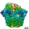 25502MC M: map data used to model this data C: citing same article ( |
|---|---|
| Similar structure data |


-Links
 PDBj
PDBj
- Assembly
| Deposited unit | 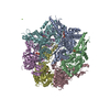
|
|---|---|
| 1 |
|
-Components
| #1: Protein | Mass: 108839.602 Da / Num. of mol.: 6 / Fragment: UNP residues 182-1133 Source method: isolated from a genetically manipulated source Source: (gene. exp.) Saccharomyces cerevisiae (strain ATCC 204508 / S288c) (yeast)Strain: ATCC 204508 / S288c / Gene: PIM1, LON, YBL022C, YBL0440 / Production host: Escherichia coli (E. coli) / References: UniProt: P36775, endopeptidase La#2: Protein/peptide | | Mass: 1039.273 Da / Num. of mol.: 1 / Source method: isolated from a natural source / Source: (natural) Escherichia coli (E. coli)#3: Chemical | ChemComp-ATP / Adenosine triphosphate  Mass: 507.181 Da / Num. of mol.: 4 / Source method: obtained synthetically / Formula: C10H16N5O13P3 / Feature type: SUBJECT OF INVESTIGATION / Comment: ATP, energy-carrying molecule*YM Mass: 507.181 Da / Num. of mol.: 4 / Source method: obtained synthetically / Formula: C10H16N5O13P3 / Feature type: SUBJECT OF INVESTIGATION / Comment: ATP, energy-carrying molecule*YM#4: Chemical | ChemComp-MG /   Mass: 24.305 Da / Num. of mol.: 4 / Source method: obtained synthetically / Formula: Mg / Feature type: SUBJECT OF INVESTIGATION Mass: 24.305 Da / Num. of mol.: 4 / Source method: obtained synthetically / Formula: Mg / Feature type: SUBJECT OF INVESTIGATION#5: Chemical | Adenosine diphosphate  Mass: 427.201 Da / Num. of mol.: 2 / Source method: obtained synthetically / Formula: C10H15N5O10P2 / Feature type: SUBJECT OF INVESTIGATION / Comment: ADP, energy-carrying molecule*YM Mass: 427.201 Da / Num. of mol.: 2 / Source method: obtained synthetically / Formula: C10H15N5O10P2 / Feature type: SUBJECT OF INVESTIGATION / Comment: ADP, energy-carrying molecule*YMHas ligand of interest | Y | |
|---|
-Experimental details
-Experiment
| Experiment | Method: ELECTRON MICROSCOPY |
|---|---|
| EM experiment | Aggregation state: PARTICLE / 3D reconstruction method: single particle reconstruction |
- Sample preparation
| Component | Name: Cryo-EM structure of yeast Lon (PIM1) in complex with endogenous substrate Type: COMPLEX / Entity ID: #1-#2 / Source: MULTIPLE SOURCES |
|---|---|
| Source (natural) | Organism: Saccharomyces cerevisiae (brewer's yeast) |
| Source (recombinant) | Organism: Escherichia coli (E. coli) |
| Buffer solution | pH: 8 |
| Specimen | Conc.: 3 mg/ml / Embedding applied: NO / Shadowing applied: NO / Staining applied: NO / Vitrification applied: YES |
| Vitrification | Cryogen name: ETHANE |
- Electron microscopy imaging
| Experimental equipment |  Model: Talos Arctica / Image courtesy: FEI Company |
|---|---|
| Microscopy | Model: FEI TALOS ARCTICA |
| Electron gun | Electron source: FIELD EMISSION GUN / Accelerating voltage: 200 kV / Illumination mode: FLOOD BEAM |
| Electron lens | Mode: BRIGHT FIELDBright-field microscopy |
| Image recording | Electron dose: 50 e/Å2 / Detector mode: COUNTING / Film or detector model: GATAN K2 SUMMIT (4k x 4k) |
- Processing
| Software | Name: PHENIX / Version: 1.19.2_4158: / Classification: refinement | ||||||||||||||||||||||||
|---|---|---|---|---|---|---|---|---|---|---|---|---|---|---|---|---|---|---|---|---|---|---|---|---|---|
| CTF correction | Type: PHASE FLIPPING ONLY | ||||||||||||||||||||||||
| 3D reconstruction | Resolution: 3.3 Å / Resolution method: FSC 0.143 CUT-OFF / Num. of particles: 81945 / Symmetry type: POINT | ||||||||||||||||||||||||
| Refine LS restraints |
|
