 Movie
Movie Controller

[English] 日本語
 Yorodumi
- PDB-7su1: Crystal structure of an acidic pH-selective Ipilimumab variant Ip... -
+ Open data

- Basic information
| Entry | Database: PDB / ID: 7su1 | ||||||
|---|---|---|---|---|---|---|---|
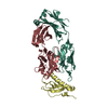
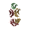
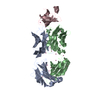
| Title | Crystal structure of an acidic pH-selective Ipilimumab variant Ipi.106 in complex with CTLA-4 | ||||||
 Components Components |
| ||||||
 Keywords Keywords |  IMMUNE SYSTEM / Immunoglobulin / checkpoint / antibody / complex IMMUNE SYSTEM / Immunoglobulin / checkpoint / antibody / complex | ||||||
| Function / homology |  Function and homology information Function and homology informationprotein complex involved in cell adhesion / negative regulation of regulatory T cell differentiation / RUNX1 and FOXP3 control the development of regulatory T lymphocytes (Tregs) / clathrin-coated endocytic vesicle / CTLA4 inhibitory signaling / negative regulation of B cell proliferation / negative regulation of T cell proliferation / B cell receptor signaling pathway / T cell receptor signaling pathway / adaptive immune response ...protein complex involved in cell adhesion / negative regulation of regulatory T cell differentiation / RUNX1 and FOXP3 control the development of regulatory T lymphocytes (Tregs) / clathrin-coated endocytic vesicle / CTLA4 inhibitory signaling / negative regulation of B cell proliferation / negative regulation of T cell proliferation / B cell receptor signaling pathway / T cell receptor signaling pathway / adaptive immune response / immune response / positive regulation of apoptotic process / external side of plasma membrane / DNA damage response / perinuclear region of cytoplasm / Golgi apparatus / plasma membraneSimilarity search - Function | ||||||
| Biological species |  Homo sapiens (human) Homo sapiens (human) | ||||||
| Method | X-RAY DIFFRACTION / SYNCHROTRON / MOLECULAR REPLACEMENT / Resolution: 2.53 Å | ||||||
 Authors Authors | Lee, P.S. / Chau, B. / Strop, P. | ||||||
| Funding support | 1items
| ||||||
 Citation Citation | Journal: Mabs / Year: 2022 Title: Improved therapeutic index of an acidic pH-selective antibody. Authors: Lee, P.S. / MacDonald, K.G. / Massi, E. / Chew, P.V. / Bee, C. / Perkins, P. / Chau, B. / Thudium, K. / Lohre, J. / Nandi, P. / Deyanova, E.G. / Barman, I. / Gudmundsson, O. / Dollinger, G. ...Authors: Lee, P.S. / MacDonald, K.G. / Massi, E. / Chew, P.V. / Bee, C. / Perkins, P. / Chau, B. / Thudium, K. / Lohre, J. / Nandi, P. / Deyanova, E.G. / Barman, I. / Gudmundsson, O. / Dollinger, G. / Sproul, T. / Engelhardt, J.J. / Strop, P. / Rajpal, A. | ||||||
| History |
|
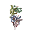
- Structure visualization
| Structure viewer | Molecule: MolmilJmol/JSmol |
|---|
- Downloads & links
-Download
| PDBx/mmCIF format | 7su1.cif.gz | 267.2 KB | Display | PDBx/mmCIF format |
|---|---|---|---|---|
| PDB format | pdb7su1.ent.gz | 180.8 KB | Display | PDB format |
| PDBx/mmJSON format | 7su1.json.gz | Tree view | PDBx/mmJSON format | |
| Others |  Other downloads Other downloads |
-Validation report
| Arichive directory | https://data.pdbj.org/pub/pdb/validation_reports/su/7su1ftp://data.pdbj.org/pub/pdb/validation_reports/su/7su1 | HTTPS FTP |
|---|
-Related structure data
| Related structure data |  7su0C  3oskS 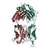 4nm4S  5truS S: Starting model for refinement C: citing same article ( |
|---|---|
| Similar structure data |


-Links
 PDBj
PDBj


- Assembly
| Deposited unit | 
| ||||||||||||
|---|---|---|---|---|---|---|---|---|---|---|---|---|---|
| 1 |
| ||||||||||||
| Unit cell |
|
-Components
| #1: Antibody | Fragment antigen-binding Mass: 24786.727 Da / Num. of mol.: 1 Source method: isolated from a genetically manipulated source Source: (gene. exp.) Homo sapiens (human) / Production host: Homo sapiens (human) |
|---|---|
| #2: Antibody | Fragment antigen-binding Mass: 23527.035 Da / Num. of mol.: 1 Source method: isolated from a genetically manipulated source Source: (gene. exp.) Homo sapiens (human) / Production host: Homo sapiens (human) |
| #3: Protein | Cytotoxic T cell / Cytotoxic T-lymphocyte-associated antigen 4 / CTLA-4 Mass: 13555.421 Da / Num. of mol.: 1 Source method: isolated from a genetically manipulated source Source: (gene. exp.) Homo sapiens (human) / Gene: CTLA4, CD152 / Production host: Homo sapiens (human) / References: UniProt: P16410 |
| #4: Sugar | ChemComp-NAG / N-Acetylglucosamine  Type: D-saccharide, beta linking / Mass: 221.208 Da / Num. of mol.: 1 / Source method: obtained synthetically / Formula: C8H15NO6 Type: D-saccharide, beta linking / Mass: 221.208 Da / Num. of mol.: 1 / Source method: obtained synthetically / Formula: C8H15NO6 |
| #5: Water | ChemComp-HOH / Water Mass: 18.015 Da / Num. of mol.: 26 / Source method: isolated from a natural source / Formula: H2O Mass: 18.015 Da / Num. of mol.: 26 / Source method: isolated from a natural source / Formula: H2O |
| Has ligand of interest | N |
-Experimental details
-Experiment
| Experiment | Method: X-RAY DIFFRACTION / Number of used crystals: 1 |
|---|
- Sample preparation
| Crystal | Density Matthews: 3.62 Å3/Da / Density % sol: 65.98 % |
|---|---|
| Crystal grow | Temperature: 293 K / Method: vapor diffusion, sitting drop Details: 0.2 M sodium citrate tribasic dehydrate, 22% PEG 3,350 |
-Data collection
| Diffraction | Mean temperature: 100 K / Serial crystal experiment: N |
|---|---|
| Diffraction source | Source: SYNCHROTRON / Site: APS  / Beamline: 17-ID / Wavelength: 1 Å / Beamline: 17-ID / Wavelength: 1 Å |
| Detector | Type: DECTRIS EIGER2 X 9M / Detector: PIXEL / Date: Oct 28, 2020 |
| Radiation | Protocol: SINGLE WAVELENGTH / Monochromatic (M) / Laue (L): M / Scattering type: x-ray |
| Radiation wavelength | Wavelength: 1 Å / Relative weight: 1 |
| Reflection | Resolution: 2.526→50 Å / Num. obs: 28083 / % possible obs: 94.6 % / Redundancy: 13.3 % / Biso Wilson estimate: 65.73 Å2 / CC1/2: 0.999 / Rmerge(I) obs: 0.101 / Rpim(I) all: 0.029 / Net I/σ(I): 15.9 |
| Reflection shell | Resolution: 2.526→2.652 Å / Mean I/σ(I) obs: 1.5 / Num. unique obs: 1404 / CC1/2: 0.64 / Rpim(I) all: 0.547 |
- Processing
| Software |
| |||||||||||||||||||||||||||||||||||||||||||||||||||||||||||||||||||||||||||||
|---|---|---|---|---|---|---|---|---|---|---|---|---|---|---|---|---|---|---|---|---|---|---|---|---|---|---|---|---|---|---|---|---|---|---|---|---|---|---|---|---|---|---|---|---|---|---|---|---|---|---|---|---|---|---|---|---|---|---|---|---|---|---|---|---|---|---|---|---|---|---|---|---|---|---|---|---|---|---|
| Refinement | Method to determine structure: MOLECULAR REPLACEMENT Starting model: 5TRU, 4NM4, 3OSK Resolution: 2.53→37.52 Å / SU ML: 0.3863 / Cross valid method: FREE R-VALUE / σ(F): 1.35 / Phase error: 29.349 Stereochemistry target values: GeoStd + Monomer Library + CDL v1.2
| |||||||||||||||||||||||||||||||||||||||||||||||||||||||||||||||||||||||||||||
| Solvent computation | Shrinkage radii: 0.9 Å / VDW probe radii: 1.11 Å / Solvent model: FLAT BULK SOLVENT MODEL | |||||||||||||||||||||||||||||||||||||||||||||||||||||||||||||||||||||||||||||
| Displacement parameters | Biso mean: 75.45 Å2 | |||||||||||||||||||||||||||||||||||||||||||||||||||||||||||||||||||||||||||||
| Refinement step | Cycle: LAST / Resolution: 2.53→37.52 Å
| |||||||||||||||||||||||||||||||||||||||||||||||||||||||||||||||||||||||||||||
| Refine LS restraints |
| |||||||||||||||||||||||||||||||||||||||||||||||||||||||||||||||||||||||||||||
| LS refinement shell |
| |||||||||||||||||||||||||||||||||||||||||||||||||||||||||||||||||||||||||||||
| Refinement TLS params. | Method: refined / Origin x: -17.3064912051 Å / Origin y: -55.6365047367 Å / Origin z: 18.4165802296 Å
| |||||||||||||||||||||||||||||||||||||||||||||||||||||||||||||||||||||||||||||
| Refinement TLS group | Selection details: all |
