 Movie
Movie Controller


+ Open data

- Basic information
| Entry | Database: PDB / ID: 7smf | ||||||
|---|---|---|---|---|---|---|---|
| Title | p107 pocket domain complexed with mutated HDAC1-3X peptide | ||||||
 Components Components |
| ||||||
 Keywords Keywords |  CELL CYCLE / Transcription / cyclin box pocket transcriptional regulator CELL CYCLE / Transcription / cyclin box pocket transcriptional regulator | ||||||
| Function / homology |  Function and homology informationregulation of lipid kinase activity / Transcription of E2F targets under negative control by p107 (RBL1) and p130 (RBL2) in complex with HDAC1 / Transcription of E2F targets under negative control by DREAM complex / negative regulation of G1/S transition of mitotic cell cycle / G1/S-Specific Transcription / negative regulation of cellular senescence / G0 and Early G1 / TP53 Regulates Transcription of Genes Involved in G2 Cell Cycle Arrest / promoter-specific chromatin binding / RNA polymerase II transcription regulatory region sequence-specific DNA binding ...regulation of lipid kinase activity / Transcription of E2F targets under negative control by p107 (RBL1) and p130 (RBL2) in complex with HDAC1 / Transcription of E2F targets under negative control by DREAM complex / negative regulation of G1/S transition of mitotic cell cycle / G1/S-Specific Transcription / negative regulation of cellular senescence / G0 and Early G1 / TP53 Regulates Transcription of Genes Involved in G2 Cell Cycle Arrest / promoter-specific chromatin binding / RNA polymerase II transcription regulatory region sequence-specific DNA binding / SMAD2/SMAD3:SMAD4 heterotrimer regulates transcription / Cyclin D associated events in G1 / chromatin organization / transcription regulator complex / cell differentiation / cell cycle / negative regulation of gene expression / chromatin / negative regulation of transcription by RNA polymerase II / nucleoplasm Function and homology informationregulation of lipid kinase activity / Transcription of E2F targets under negative control by p107 (RBL1) and p130 (RBL2) in complex with HDAC1 / Transcription of E2F targets under negative control by DREAM complex / negative regulation of G1/S transition of mitotic cell cycle / G1/S-Specific Transcription / negative regulation of cellular senescence / G0 and Early G1 / TP53 Regulates Transcription of Genes Involved in G2 Cell Cycle Arrest / promoter-specific chromatin binding / RNA polymerase II transcription regulatory region sequence-specific DNA binding ...regulation of lipid kinase activity / Transcription of E2F targets under negative control by p107 (RBL1) and p130 (RBL2) in complex with HDAC1 / Transcription of E2F targets under negative control by DREAM complex / negative regulation of G1/S transition of mitotic cell cycle / G1/S-Specific Transcription / negative regulation of cellular senescence / G0 and Early G1 / TP53 Regulates Transcription of Genes Involved in G2 Cell Cycle Arrest / promoter-specific chromatin binding / RNA polymerase II transcription regulatory region sequence-specific DNA binding / SMAD2/SMAD3:SMAD4 heterotrimer regulates transcription / Cyclin D associated events in G1 / chromatin organization / transcription regulator complex / cell differentiation / cell cycle / negative regulation of gene expression / chromatin / negative regulation of transcription by RNA polymerase II / nucleoplasmSimilarity search - Function | ||||||
| Biological species |  Homo sapiens (human) Homo sapiens (human) | ||||||
| Method | X-RAY DIFFRACTION / SYNCHROTRON / MOLECULAR REPLACEMENT / Resolution: 3 Å | ||||||
 Authors Authors | Putta, S. / Fernandez, S.M. / Tripathi, S.M. / Muller, G.A. / Rubin, S.M. | ||||||
| Funding support |  United States, 1items United States, 1items
| ||||||
 Citation Citation | Journal: Structure / Year: 2022 Title: Structural basis for tunable affinity and specificity of LxCxE-dependent protein interactions with the retinoblastoma protein family. Authors: Putta, S. / Alvarez, L. / Ludtke, S. / Sehr, P. / Muller, G.A. / Fernandez, S.M. / Tripathi, S. / Lewis, J. / Gibson, T.J. / Chemes, L.B. / Rubin, S.M. | ||||||
| History |
|
- Structure visualization
| Structure viewer | Molecule: MolmilJmol/JSmol |
|---|
- Downloads & links
-Download
| PDBx/mmCIF format | 7smf.cif.gz | 298.5 KB | Display | PDBx/mmCIF format |
|---|---|---|---|---|
| PDB format | pdb7smf.ent.gz | 240.2 KB | Display | PDB format |
| PDBx/mmJSON format | 7smf.json.gz | Tree view | PDBx/mmJSON format | |
| Others |  Other downloads Other downloads |
-Validation report
| Arichive directory | https://data.pdbj.org/pub/pdb/validation_reports/sm/7smfftp://data.pdbj.org/pub/pdb/validation_reports/sm/7smf | HTTPS FTP |
|---|
-Related structure data
| Related structure data |  7smcC 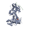 7smdC 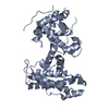 7smeC 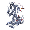 4yosS S: Starting model for refinement C: citing same article ( |
|---|---|
| Similar structure data |
-Links
 PDBj
PDBj


- Assembly
| Deposited unit | 
| ||||||||
|---|---|---|---|---|---|---|---|---|---|
| 1 | 
| ||||||||
| 2 | 
| ||||||||
| Unit cell |
|
-Components
| #1: Protein | / 107 kDa retinoblastoma-associated protein / p107 / pRb1 Mass: 43265.965 Da / Num. of mol.: 2 / Fragment: UNP residues 391-601,780-887,924-972 Source method: isolated from a genetically manipulated source Source: (gene. exp.) Homo sapiens (human) / Gene: RBL1 / Production host:  Escherichia coli (E. coli) / References: UniProt: P28749 Escherichia coli (E. coli) / References: UniProt: P28749#2: Protein/peptide | HDAC1Mass: 1283.318 Da / Num. of mol.: 2 / Fragment: UNP residues 413-422 / Mutation: R413D, A415Y, E417Y / Source method: obtained synthetically / Source: (synth.) Homo sapiens (human)References: histone deacetylase, Hydrolases; Acting on carbon-nitrogen bonds, other than peptide bonds; In linear amides#3: Chemical | ChemComp-SO4 / Sulfate  Mass: 96.063 Da / Num. of mol.: 4 / Source method: obtained synthetically / Formula: SO4 Mass: 96.063 Da / Num. of mol.: 4 / Source method: obtained synthetically / Formula: SO4#4: Water | ChemComp-HOH / | Water Mass: 18.015 Da / Num. of mol.: 41 / Source method: isolated from a natural source / Formula: H2O Mass: 18.015 Da / Num. of mol.: 41 / Source method: isolated from a natural source / Formula: H2OHas ligand of interest | N | |
|---|
-Experimental details
-Experiment
| Experiment | Method: X-RAY DIFFRACTION / Number of used crystals: 1 |
|---|
- Sample preparation
| Crystal | Density Matthews: 3.34 Å3/Da / Density % sol: 63.19 % |
|---|---|
| Crystal grow | Temperature: 277 K / Method: vapor diffusion, hanging drop Details: 100 mM MES, pH 6.5, 4% PEG400, 1.6 M ammonium sulfate |
-Data collection
| Diffraction | Mean temperature: 100 K / Serial crystal experiment: N |
|---|---|
| Diffraction source | Source: SYNCHROTRON / Site: APS / Beamline: 23-ID-D / Wavelength: 1.033 Å |
| Detector | Type: DECTRIS PILATUS 6M / Detector: PIXEL / Date: Jul 27, 2021 |
| Radiation | Protocol: SINGLE WAVELENGTH / Monochromatic (M) / Laue (L): M / Scattering type: x-ray |
| Radiation wavelength | Wavelength: 1.033 Å / Relative weight: 1 |
| Reflection | Resolution: 3→39.29 Å / Num. obs: 21589 / % possible obs: 93.5 % / Redundancy: 2 % / CC1/2: 0.81 / Rmerge(I) obs: 0.251 / Net I/σ(I): 3.1 |
| Reflection shell | Resolution: 3→3.18 Å / Rmerge(I) obs: 0.74 / Mean I/σ(I) obs: 1.3 / Num. unique obs: 3425 / CC1/2: 0.64 |
- Processing
| Software |
| ||||||||||||||||||||||||||||||||||||||||||||||||||||||||||||||||||||||||||||||||||||||||||||||||||||||||||||||||||||||||||||||||||||||||||||||||||||||||||||||||||||||||||||||||||||||||||||||||||||||||
|---|---|---|---|---|---|---|---|---|---|---|---|---|---|---|---|---|---|---|---|---|---|---|---|---|---|---|---|---|---|---|---|---|---|---|---|---|---|---|---|---|---|---|---|---|---|---|---|---|---|---|---|---|---|---|---|---|---|---|---|---|---|---|---|---|---|---|---|---|---|---|---|---|---|---|---|---|---|---|---|---|---|---|---|---|---|---|---|---|---|---|---|---|---|---|---|---|---|---|---|---|---|---|---|---|---|---|---|---|---|---|---|---|---|---|---|---|---|---|---|---|---|---|---|---|---|---|---|---|---|---|---|---|---|---|---|---|---|---|---|---|---|---|---|---|---|---|---|---|---|---|---|---|---|---|---|---|---|---|---|---|---|---|---|---|---|---|---|---|---|---|---|---|---|---|---|---|---|---|---|---|---|---|---|---|---|---|---|---|---|---|---|---|---|---|---|---|---|---|---|---|---|
| Refinement | Method to determine structure: MOLECULAR REPLACEMENT Starting model: PDB entry 4YOS Resolution: 3→39.29 Å / SU ML: 0.49 / Cross valid method: FREE R-VALUE / σ(F): 1.96 / Phase error: 30.8 / Stereochemistry target values: ML
| ||||||||||||||||||||||||||||||||||||||||||||||||||||||||||||||||||||||||||||||||||||||||||||||||||||||||||||||||||||||||||||||||||||||||||||||||||||||||||||||||||||||||||||||||||||||||||||||||||||||||
| Solvent computation | Shrinkage radii: 0.9 Å / VDW probe radii: 1.11 Å / Solvent model: FLAT BULK SOLVENT MODEL | ||||||||||||||||||||||||||||||||||||||||||||||||||||||||||||||||||||||||||||||||||||||||||||||||||||||||||||||||||||||||||||||||||||||||||||||||||||||||||||||||||||||||||||||||||||||||||||||||||||||||
| Refinement step | Cycle: LAST / Resolution: 3→39.29 Å
| ||||||||||||||||||||||||||||||||||||||||||||||||||||||||||||||||||||||||||||||||||||||||||||||||||||||||||||||||||||||||||||||||||||||||||||||||||||||||||||||||||||||||||||||||||||||||||||||||||||||||
| Refine LS restraints |
| ||||||||||||||||||||||||||||||||||||||||||||||||||||||||||||||||||||||||||||||||||||||||||||||||||||||||||||||||||||||||||||||||||||||||||||||||||||||||||||||||||||||||||||||||||||||||||||||||||||||||
| LS refinement shell |
| ||||||||||||||||||||||||||||||||||||||||||||||||||||||||||||||||||||||||||||||||||||||||||||||||||||||||||||||||||||||||||||||||||||||||||||||||||||||||||||||||||||||||||||||||||||||||||||||||||||||||
| Refinement TLS params. | Method: refined / Refine-ID: X-RAY DIFFRACTION
| ||||||||||||||||||||||||||||||||||||||||||||||||||||||||||||||||||||||||||||||||||||||||||||||||||||||||||||||||||||||||||||||||||||||||||||||||||||||||||||||||||||||||||||||||||||||||||||||||||||||||
| Refinement TLS group |
|
