Entry Database : PDB / ID : 7siyTitle cCBL TKB domain in complex with pZAP70 peptide E3 ubiquitin-protein ligase CBL Peptide from Tyrosine-protein kinase ZAP-70 Keywords / / / Function / homology Function Domain/homology Component
/ / / / / / / / / / / / / / / / / / / / / / / / / / / / / / / / / / / / / / / / / / / / / / / / / / / / / / / / / / / / / / / / / / / / / / / / / / / / / / / / / / / / / / / / / / / / / / / / / / / / / / / / / / / / / / / / / / / / / / / / / / / / / / / / / / / / / / / / / / / / / / / / / / / / / / / / / / / / / / / Biological species Homo sapiens (human)Method / / / Resolution : 1.48 Å Authors Murray, J.M. / Yu, C. Funding support 1items Organization Grant number Country Not funded
Journal : To Be Published Title : cCBL TKB domain in complex with pZAP70 peptideAuthors : Murray, J.M. / Yu, C. History Deposition Oct 15, 2021 Deposition site / Processing site Revision 1.0 Nov 9, 2022 Provider / Type Revision 1.1 Oct 18, 2023 Group / Refinement descriptionCategory / chem_comp_bond / pdbx_initial_refinement_modelRevision 1.2 Nov 15, 2023 Group / Category / chem_comp_bond / Item / _chem_comp_bond.atom_id_2
Show all Show less
 Movie
Movie Controller


 Open data
 Basic information
 Components
 Keywords
 IMMUNE SYSTEM / cCBL /
  Function and homology information
 Authors
 Citation
 Structure visualization
 Downloads & links
 Other downloads
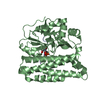
 PDBj
PDBj


 Assembly


 Mass: 24.305 Da / Num. of mol.: 1 / Source method: obtained synthetically / Formula: Mg
 Mass: 18.015 Da / Num. of mol.: 372 / Source method: isolated from a natural source / Formula: H2O
 Sample preparation
 / Beamline: BL12-2 / Wavelength: 0.97946 Å
 Processing