 Movie
Movie Controller

[English] 日本語
 Yorodumi
- PDB-7s6t: Complex structure of Methane monooxygenase hydroxylase and regula... -
+ Open data

- Basic information
| Entry | Database: PDB / ID: 7s6t | ||||||||||||
|---|---|---|---|---|---|---|---|---|---|---|---|---|---|
| Title | Complex structure of Methane monooxygenase hydroxylase and regulatory subunit H33A | ||||||||||||
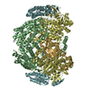
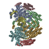
 Components Components | (Methane monooxygenase ... ) x 4 ) x 4 | ||||||||||||
 Keywords Keywords | HYDROLASE / OXIDOREDUCTASE | ||||||||||||
| Function / homology |  Function and homology informationmethane monooxygenase activity / methane metabolic process / oxidoreductase activity, acting on paired donors, with incorporation or reduction of molecular oxygen, NAD(P)H as one donor, and incorporation of one atom of oxygen / : / monooxygenase activity Function and homology informationmethane monooxygenase activity / methane metabolic process / oxidoreductase activity, acting on paired donors, with incorporation or reduction of molecular oxygen, NAD(P)H as one donor, and incorporation of one atom of oxygen / : / monooxygenase activitySimilarity search - Function | ||||||||||||
| Biological species |  Methylosinus trichosporium OB3b (bacteria) Methylosinus trichosporium OB3b (bacteria) | ||||||||||||
| Method | X-RAY DIFFRACTION / SYNCHROTRON / FOURIER SYNTHESIS / Resolution: 1.82 Å | ||||||||||||
 Authors Authors | Johns, J.C. / Banerjee, R. / Semonis, M.M. / Shi, K. / Aihara, H. / Lipscomb, J.D. | ||||||||||||
| Funding support |  United States, 3items United States, 3items
| ||||||||||||
 Citation Citation | Journal: Biochemistry / Year: 2022 Title: X-ray Crystal Structures of Methane Monooxygenase Hydroxylase Complexes with Variants of Its Regulatory Component: Correlations with Altered Reaction Cycle Dynamics. Authors: Jones, J.C. / Banerjee, R. / Semonis, M.M. / Shi, K. / Aihara, H. / Lipscomb, J.D. | ||||||||||||
| History |
|
- Structure visualization
| Structure viewer | Molecule: MolmilJmol/JSmol |
|---|
- Downloads & links
-Download
| PDBx/mmCIF format | 7s6t.cif.gz | 548.7 KB | Display | PDBx/mmCIF format |
|---|---|---|---|---|
| PDB format | pdb7s6t.ent.gz | 437.5 KB | Display | PDB format |
| PDBx/mmJSON format | 7s6t.json.gz | Tree view | PDBx/mmJSON format | |
| Others |  Other downloads Other downloads |
-Validation report
| Arichive directory | https://data.pdbj.org/pub/pdb/validation_reports/s6/7s6tftp://data.pdbj.org/pub/pdb/validation_reports/s6/7s6t | HTTPS FTP |
|---|
-Related structure data
| Related structure data | 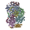 7s6qC  7s6rC  7s6sC  7s7hC  7m8qS S: Starting model for refinement C: citing same article ( |
|---|---|
| Similar structure data |


-Links
 PDBj
PDBj
- Assembly
| Deposited unit | 
| |||||||||||||||||||||||||||||||||||||||||||||||||||||||||||||||||||||||||
|---|---|---|---|---|---|---|---|---|---|---|---|---|---|---|---|---|---|---|---|---|---|---|---|---|---|---|---|---|---|---|---|---|---|---|---|---|---|---|---|---|---|---|---|---|---|---|---|---|---|---|---|---|---|---|---|---|---|---|---|---|---|---|---|---|---|---|---|---|---|---|---|---|---|---|
| 1 |
| |||||||||||||||||||||||||||||||||||||||||||||||||||||||||||||||||||||||||
| Unit cell |
| |||||||||||||||||||||||||||||||||||||||||||||||||||||||||||||||||||||||||
| Noncrystallographic symmetry (NCS) | NCS domain:
NCS domain segments:
|