 Movie
Movie Controller


+ Open data

- Basic information
| Entry | Database: PDB / ID: 7rsw | ||||||
|---|---|---|---|---|---|---|---|
| Title | Crystal structure of group B human rotavirus VP8* | ||||||
 Components Components |
| ||||||
 Keywords Keywords |  STRUCTURAL PROTEIN / Rotavirus / capsid / glycan-binding STRUCTURAL PROTEIN / Rotavirus / capsid / glycan-binding | ||||||
| Function / homology |  Function and homology information Function and homology informationhost cell rough endoplasmic reticulum / viral outer capsid / permeabilization of host organelle membrane involved in viral entry into host cell / symbiont entry into host cell via permeabilization of inner membrane / host cell endoplasmic reticulum-Golgi intermediate compartment / virion attachment to host cell / host cell plasma membrane / membraneSimilarity search - Function | ||||||
| Biological species |  Rotavirus BRotavirus Rotavirus BRotavirus | ||||||
| Method | X-RAY DIFFRACTION / SYNCHROTRON / AB INITIO PHASING / Resolution: 1.32 Å | ||||||
 Authors Authors | Hu, L. / Salmen, W. / Sankaran, B. / Prasad, B.V. | ||||||
| Funding support |  United States, 1items United States, 1items
| ||||||
 Citation Citation | Journal: Commun Biol / Year: 2022 Title: Novel fold of rotavirus glycan-binding domain predicted by AlphaFold2 and determined by X-ray crystallography. Authors: Hu, L. / Salmen, W. / Sankaran, B. / Lasanajak, Y. / Smith, D.F. / Crawford, S.E. / Estes, M.K. / Prasad, B.V.V. | ||||||
| History |
|
- Structure visualization
| Structure viewer | Molecule: MolmilJmol/JSmol |
|---|
- Downloads & links
-Download
| PDBx/mmCIF format | 7rsw.cif.gz | 132.2 KB | Display | PDBx/mmCIF format |
|---|---|---|---|---|
| PDB format | pdb7rsw.ent.gz | 86.3 KB | Display | PDB format |
| PDBx/mmJSON format | 7rsw.json.gz | Tree view | PDBx/mmJSON format | |
| Others |  Other downloads Other downloads |
-Validation report
| Arichive directory | https://data.pdbj.org/pub/pdb/validation_reports/rs/7rswftp://data.pdbj.org/pub/pdb/validation_reports/rs/7rsw | HTTPS FTP |
|---|
-Related structure data
| Similar structure data |
|---|
-Links
 PDBj
PDBj- Assembly
| Deposited unit | 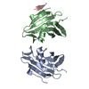
| ||||||||||||
|---|---|---|---|---|---|---|---|---|---|---|---|---|---|
| 1 | 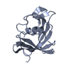
| ||||||||||||
| 2 | 
| ||||||||||||
| 3 | 
| ||||||||||||
| Unit cell |
|
-Components
| #1: Protein | Mass: 18610.883 Da / Num. of mol.: 2 Source method: isolated from a genetically manipulated source Source: (gene. exp.) Rotavirus B (isolate RVB/Human/China/ADRV/1982)Strain: isolate RVB/Human/China/ADRV/1982 / Gene: VP4 / Production host:  Escherichia coli BL21(DE3) (bacteria) / References: UniProt: H0USR8 Escherichia coli BL21(DE3) (bacteria) / References: UniProt: H0USR8#2: Protein/peptide | | Mass: 274.274 Da / Num. of mol.: 1 / Source method: isolated from a natural source / Source: (natural) Rotavirus#3: Water | ChemComp-HOH / | Water Mass: 18.015 Da / Num. of mol.: 285 / Source method: isolated from a natural source / Formula: H2O Mass: 18.015 Da / Num. of mol.: 285 / Source method: isolated from a natural source / Formula: H2O |
|---|
-Experimental details
-Experiment
| Experiment | Method: X-RAY DIFFRACTION / Number of used crystals: 1 |
|---|
- Sample preparation
| Crystal | Density Matthews: 2.01 Å3/Da / Density % sol: 38.74 % |
|---|---|
| Crystal grow | Temperature: 293 K / Method: vapor diffusion, hanging drop Details: 0.2 M ammonium sulfate, 30% PEG 2000 MME, 0.1 M sodium acetate, pH 4.6 |
-Data collection
| Diffraction | Mean temperature: 100 K / Serial crystal experiment: N |
|---|---|
| Diffraction source | Source: SYNCHROTRON / Site: ALS / Beamline: 5.0.1 / Wavelength: 0.97741 Å |
| Detector | Type: DECTRIS PILATUS 6M / Detector: PIXEL / Date: Aug 14, 2018 |
| Radiation | Protocol: SINGLE WAVELENGTH / Monochromatic (M) / Laue (L): M / Scattering type: x-ray |
| Radiation wavelength | Wavelength: 0.97741 Å / Relative weight: 1 |
| Reflection | Resolution: 1.32→30.41 Å / Num. obs: 41757 / % possible obs: 82.69 % / Redundancy: 1.9 % / Biso Wilson estimate: 13.24 Å2 / Rmerge(I) obs: 0.04 / Net I/σ(I): 19.03 |
| Reflection shell | Resolution: 1.32→1.34 Å / Rmerge(I) obs: 0.262 / Mean I/σ(I) obs: 2.31 / Num. unique obs: 1675 |
- Processing
| Software |
| |||||||||||||||||||||||||||||||||||||||||||||||||||||||||||||||||||||||||||||||||||||||||||||||||||||||||
|---|---|---|---|---|---|---|---|---|---|---|---|---|---|---|---|---|---|---|---|---|---|---|---|---|---|---|---|---|---|---|---|---|---|---|---|---|---|---|---|---|---|---|---|---|---|---|---|---|---|---|---|---|---|---|---|---|---|---|---|---|---|---|---|---|---|---|---|---|---|---|---|---|---|---|---|---|---|---|---|---|---|---|---|---|---|---|---|---|---|---|---|---|---|---|---|---|---|---|---|---|---|---|---|---|---|---|
| Refinement | Method to determine structure: AB INITIO PHASING / Resolution: 1.32→30.41 Å / SU ML: 0.1577 / Cross valid method: FREE R-VALUE / σ(F): 1.36 / Phase error: 22.1923 Stereochemistry target values: GeoStd + Monomer Library + CDL v1.2
| |||||||||||||||||||||||||||||||||||||||||||||||||||||||||||||||||||||||||||||||||||||||||||||||||||||||||
| Solvent computation | Shrinkage radii: 0.9 Å / VDW probe radii: 1.11 Å / Solvent model: FLAT BULK SOLVENT MODEL | |||||||||||||||||||||||||||||||||||||||||||||||||||||||||||||||||||||||||||||||||||||||||||||||||||||||||
| Displacement parameters | Biso mean: 19.2 Å2 | |||||||||||||||||||||||||||||||||||||||||||||||||||||||||||||||||||||||||||||||||||||||||||||||||||||||||
| Refinement step | Cycle: LAST / Resolution: 1.32→30.41 Å
| |||||||||||||||||||||||||||||||||||||||||||||||||||||||||||||||||||||||||||||||||||||||||||||||||||||||||
| Refine LS restraints |
| |||||||||||||||||||||||||||||||||||||||||||||||||||||||||||||||||||||||||||||||||||||||||||||||||||||||||
| LS refinement shell |
|
