 Movie
Movie Controller

[English] 日本語
 Yorodumi
- PDB-7rom: Crystal structure of Saccharomyces cerevisiae NADH-cytochrome b5 ... -
+ Open data

- Basic information
| Entry | Database: PDB / ID: 7rom | |||||||||||||||
|---|---|---|---|---|---|---|---|---|---|---|---|---|---|---|---|---|
| Title | Crystal structure of Saccharomyces cerevisiae NADH-cytochrome b5 reductase 1 (Cbr1) fragment (residues 28-284) bound to FAD | |||||||||||||||
 Components Components | NADH-cytochrome b5 reductase 1 | |||||||||||||||
 Keywords Keywords |  OXIDOREDUCTASE / Reductase / electron transfer OXIDOREDUCTASE / Reductase / electron transfer | |||||||||||||||
| Function / homology |  Function and homology information Function and homology informationVitamin C (ascorbate) metabolism / cytochrome-b5 reductase / tRNA wobble base 5-methoxycarbonylmethyl-2-thiouridinylation / cytochrome-b5 reductase activity, acting on NAD(P)H / protein histidyl modification to diphthamide / Platelet degranulation / NADH dehydrogenase activity / Neutrophil degranulation / mitochondrial outer membrane / membrane => GO:0016020 ...Vitamin C (ascorbate) metabolism / cytochrome-b5 reductase / tRNA wobble base 5-methoxycarbonylmethyl-2-thiouridinylation / cytochrome-b5 reductase activity, acting on NAD(P)H / protein histidyl modification to diphthamide / Platelet degranulation / NADH dehydrogenase activity / Neutrophil degranulation / mitochondrial outer membrane / membrane => GO:0016020 / endoplasmic reticulum membrane / mitochondrion / nucleus / plasma membraneSimilarity search - Function | |||||||||||||||
| Biological species |  Saccharomyces cerevisiae (brewer's yeast) Saccharomyces cerevisiae (brewer's yeast) | |||||||||||||||
| Method | X-RAY DIFFRACTION / SYNCHROTRON / MOLECULAR REPLACEMENT / Resolution: 1.65 Å | |||||||||||||||
 Authors Authors | Fenwick, M.K. / Zhang, Y. / Lin, H. | |||||||||||||||
| Funding support |  United States, 4items United States, 4items
| |||||||||||||||
 Citation Citation | Journal: To Be Published Title: Crystal structure of Saccharomyces cerevisiae NADH-cytochrome b5 reductase 1 (Cbr1) fragment (residues 28-284) bound to FAD Authors: Fenwick, M.K. / Lin, H. | |||||||||||||||
| History |
|
- Structure visualization
| Structure viewer | Molecule: MolmilJmol/JSmol |
|---|
- Downloads & links
-Download
| PDBx/mmCIF format | 7rom.cif.gz | 135.3 KB | Display | PDBx/mmCIF format |
|---|---|---|---|---|
| PDB format | pdb7rom.ent.gz | 101.9 KB | Display | PDB format |
| PDBx/mmJSON format | 7rom.json.gz | Tree view | PDBx/mmJSON format | |
| Others |  Other downloads Other downloads |
-Validation report
| Arichive directory | https://data.pdbj.org/pub/pdb/validation_reports/ro/7romftp://data.pdbj.org/pub/pdb/validation_reports/ro/7rom | HTTPS FTP |
|---|
-Related structure data
| Related structure data | 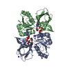 2eixS S: Starting model for refinement |
|---|---|
| Similar structure data |
-Links
 PDBj
PDBj


- Assembly
| Deposited unit | 
| ||||||||
|---|---|---|---|---|---|---|---|---|---|
| 1 |
| ||||||||
| Unit cell |
|
-Components
| #1: Protein | Mass: 29029.584 Da / Num. of mol.: 1 Source method: isolated from a genetically manipulated source Source: (gene. exp.) Saccharomyces cerevisiae (brewer's yeast)Gene: CBR1, CBR, CBR5, YIL043C / Production host:  Escherichia coli BL21(DE3) (bacteria) / References: UniProt: P38626, cytochrome-b5 reductase Escherichia coli BL21(DE3) (bacteria) / References: UniProt: P38626, cytochrome-b5 reductase | ||||||
|---|---|---|---|---|---|---|---|
| #2: Chemical | ChemComp-FAD / Flavin adenine dinucleotide  Mass: 785.550 Da / Num. of mol.: 1 / Source method: obtained synthetically / Formula: C27H33N9O15P2 / Feature type: SUBJECT OF INVESTIGATION / Comment: FAD*YM Mass: 785.550 Da / Num. of mol.: 1 / Source method: obtained synthetically / Formula: C27H33N9O15P2 / Feature type: SUBJECT OF INVESTIGATION / Comment: FAD*YM | ||||||
| #3: Chemical | Chloride  Mass: 35.453 Da / Num. of mol.: 3 / Source method: obtained synthetically / Formula: Cl Mass: 35.453 Da / Num. of mol.: 3 / Source method: obtained synthetically / Formula: Cl#4: Chemical | Polyethylene glycol  Mass: 150.173 Da / Num. of mol.: 2 / Source method: obtained synthetically / Formula: C6H14O4 Mass: 150.173 Da / Num. of mol.: 2 / Source method: obtained synthetically / Formula: C6H14O4#5: Water | ChemComp-HOH / | Water Mass: 18.015 Da / Num. of mol.: 435 / Source method: isolated from a natural source / Formula: H2O Mass: 18.015 Da / Num. of mol.: 435 / Source method: isolated from a natural source / Formula: H2OHas ligand of interest | Y | |
-Experimental details
-Experiment
| Experiment | Method: X-RAY DIFFRACTION / Number of used crystals: 1 |
|---|
- Sample preparation
| Crystal | Density Matthews: 2.63 Å3/Da / Density % sol: 53.3 % |
|---|---|
| Crystal grow | Temperature: 295 K / Method: vapor diffusion / Details: 20 % (w/v) PEG 3350 and 200 mM sodium thiocyanate |
-Data collection
| Diffraction | Mean temperature: 100 K / Serial crystal experiment: N | ||||||||||||||||||||||||||||||
|---|---|---|---|---|---|---|---|---|---|---|---|---|---|---|---|---|---|---|---|---|---|---|---|---|---|---|---|---|---|---|---|
| Diffraction source | Source: SYNCHROTRON / Site: APS / Beamline: 24-ID-C / Wavelength: 0.97903 Å | ||||||||||||||||||||||||||||||
| Detector | Type: DECTRIS EIGER2 X 16M / Detector: PIXEL / Date: Jul 18, 2020 | ||||||||||||||||||||||||||||||
| Radiation | Protocol: SINGLE WAVELENGTH / Monochromatic (M) / Laue (L): M / Scattering type: x-ray | ||||||||||||||||||||||||||||||
| Radiation wavelength | Wavelength: 0.97903 Å / Relative weight: 1 | ||||||||||||||||||||||||||||||
| Reflection | Resolution: 1.65→46.65 Å / Num. obs: 36978 / % possible obs: 99.6 % / Redundancy: 8.7 % / Biso Wilson estimate: 23.44 Å2 / CC1/2: 0.999 / Rmerge(I) obs: 0.052 / Rpim(I) all: 0.018 / Rrim(I) all: 0.055 / Net I/σ(I): 21.7 / Num. measured all: 322326 / Scaling rejects: 3 | ||||||||||||||||||||||||||||||
| Reflection shell | Diffraction-ID: 1
|
- Processing
| Software |
| |||||||||||||||||||||||||||||||||||||||||||||||||||||||||||||||||||||||||||||||||||||||||||||||||||||||||||||||||||||||||||||
|---|---|---|---|---|---|---|---|---|---|---|---|---|---|---|---|---|---|---|---|---|---|---|---|---|---|---|---|---|---|---|---|---|---|---|---|---|---|---|---|---|---|---|---|---|---|---|---|---|---|---|---|---|---|---|---|---|---|---|---|---|---|---|---|---|---|---|---|---|---|---|---|---|---|---|---|---|---|---|---|---|---|---|---|---|---|---|---|---|---|---|---|---|---|---|---|---|---|---|---|---|---|---|---|---|---|---|---|---|---|---|---|---|---|---|---|---|---|---|---|---|---|---|---|---|---|---|
| Refinement | Method to determine structure: MOLECULAR REPLACEMENT Starting model: 2EIX Resolution: 1.65→46.65 Å / SU ML: 0.19 / Cross valid method: THROUGHOUT / σ(F): 1.35 / Phase error: 18.54 / Stereochemistry target values: ML
| |||||||||||||||||||||||||||||||||||||||||||||||||||||||||||||||||||||||||||||||||||||||||||||||||||||||||||||||||||||||||||||
| Solvent computation | Shrinkage radii: 0.9 Å / VDW probe radii: 1.11 Å / Solvent model: FLAT BULK SOLVENT MODEL | |||||||||||||||||||||||||||||||||||||||||||||||||||||||||||||||||||||||||||||||||||||||||||||||||||||||||||||||||||||||||||||
| Displacement parameters | Biso max: 101.12 Å2 / Biso mean: 26.8104 Å2 / Biso min: 12.06 Å2 | |||||||||||||||||||||||||||||||||||||||||||||||||||||||||||||||||||||||||||||||||||||||||||||||||||||||||||||||||||||||||||||
| Refinement step | Cycle: final / Resolution: 1.65→46.65 Å
| |||||||||||||||||||||||||||||||||||||||||||||||||||||||||||||||||||||||||||||||||||||||||||||||||||||||||||||||||||||||||||||
| LS refinement shell | Refine-ID: X-RAY DIFFRACTION / Rfactor Rfree error: 0 / Total num. of bins used: 13
| |||||||||||||||||||||||||||||||||||||||||||||||||||||||||||||||||||||||||||||||||||||||||||||||||||||||||||||||||||||||||||||
| Refinement TLS params. | Method: refined / Refine-ID: X-RAY DIFFRACTION
| |||||||||||||||||||||||||||||||||||||||||||||||||||||||||||||||||||||||||||||||||||||||||||||||||||||||||||||||||||||||||||||
| Refinement TLS group |
|
