 Movie
Movie Controller

[English] 日本語
 Yorodumi
- PDB-7rlt: Structure of ligand-free ALDH1L1 (10-formyltetrahydrofolate dehyd... -
+ Open data

- Basic information
| Entry | Database: PDB / ID: 7rlt | ||||||
|---|---|---|---|---|---|---|---|
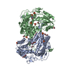
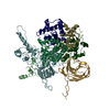
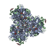
| Title | Structure of ligand-free ALDH1L1 (10-formyltetrahydrofolate dehydrogenase) | ||||||
 Components Components | Cytosolic 10-formyltetrahydrofolate dehydrogenase | ||||||
 Keywords Keywords |  CYTOSOLIC PROTEIN / folate metabolism / acyl carrier protein / peptidyl carrier protein CYTOSOLIC PROTEIN / folate metabolism / acyl carrier protein / peptidyl carrier protein | ||||||
| Function / homology |  Function and homology information Function and homology informationMetabolism of folate and pterines / aldehyde dehydrogenase (NADP+) activity / formyltetrahydrofolate dehydrogenase / formyltetrahydrofolate dehydrogenase activity / 10-formyltetrahydrofolate catabolic process / aldehyde dehydrogenase [NAD(P)+] activity / NADPH regeneration / aldehyde dehydrogenase (NAD+) activity / bile acid signaling pathway / folic acid metabolic process ...Metabolism of folate and pterines / aldehyde dehydrogenase (NADP+) activity / formyltetrahydrofolate dehydrogenase / formyltetrahydrofolate dehydrogenase activity / 10-formyltetrahydrofolate catabolic process / aldehyde dehydrogenase [NAD(P)+] activity / NADPH regeneration / aldehyde dehydrogenase (NAD+) activity / bile acid signaling pathway / folic acid metabolic process / tetrahydrofolate biosynthetic process / one-carbon metabolic process / protein-containing complex binding / protein-containing complex / cytosolSimilarity search - Function | ||||||
| Biological species |  Rattus norvegicus (Norway rat) Rattus norvegicus (Norway rat) | ||||||
| Method | ELECTRON MICROSCOPY / single particle reconstruction / cryo EM / Resolution: 3.7 Å | ||||||
 Authors Authors | Tsybovsky, Y. / Sereda, V. / Golczak, M. / Krupenko, N.I. / Krupenko, S.A. | ||||||
| Funding support |  United States, 1items United States, 1items
| ||||||
 Citation Citation | Journal: Commun Biol / Year: 2022 Title: Structure of putative tumor suppressor ALDH1L1. Authors: Yaroslav Tsybovsky / Valentin Sereda / Marcin Golczak / Natalia I Krupenko / Sergey A Krupenko / Abstract: Putative tumor suppressor ALDH1L1, the product of natural fusion of three unrelated genes, regulates folate metabolism by catalyzing NADP-dependent conversion of 10-formyltetrahydrofolate to ...Putative tumor suppressor ALDH1L1, the product of natural fusion of three unrelated genes, regulates folate metabolism by catalyzing NADP-dependent conversion of 10-formyltetrahydrofolate to tetrahydrofolate and CO. Cryo-EM structures of tetrameric rat ALDH1L1 revealed the architecture and functional domain interactions of this complex enzyme. Highly mobile N-terminal domains, which remove formyl from 10-formyltetrahydrofolate, undergo multiple transient inter-domain interactions. The C-terminal aldehyde dehydrogenase domains, which convert formyl to CO, form unusually large interfaces with the intermediate domains, homologs of acyl/peptidyl carrier proteins (A/PCPs), which transfer the formyl group between the catalytic domains. The 4'-phosphopantetheine arm of the intermediate domain is fully extended and reaches deep into the catalytic pocket of the C-terminal domain. Remarkably, the tetrameric state of ALDH1L1 is indispensable for catalysis because the intermediate domain transfers formyl between the catalytic domains of different protomers. These findings emphasize the versatility of A/PCPs in complex, highly dynamic enzymatic systems. | ||||||
| History |
|
- Structure visualization
| Movie |
Movie viewer |
|---|---|
| Structure viewer | Molecule: MolmilJmol/JSmol |
- Downloads & links
-Download
| PDBx/mmCIF format | 7rlt.cif.gz | 461.5 KB | Display | PDBx/mmCIF format |
|---|---|---|---|---|
| PDB format | pdb7rlt.ent.gz | 372.6 KB | Display | PDB format |
| PDBx/mmJSON format | 7rlt.json.gz | Tree view | PDBx/mmJSON format | |
| Others |  Other downloads Other downloads |
-Validation report
| Arichive directory | https://data.pdbj.org/pub/pdb/validation_reports/rl/7rltftp://data.pdbj.org/pub/pdb/validation_reports/rl/7rlt | HTTPS FTP |
|---|
-Related structure data
| Related structure data |  24540MC  7rluC M: map data used to model this data C: citing same article ( |
|---|---|
| Similar structure data |


-Links
 PDBj
PDBj


- Assembly
| Deposited unit | 
|
|---|---|
| 1 |
|
-Components
| #1: Protein | Mass: 98985.055 Da / Num. of mol.: 4 Source method: isolated from a genetically manipulated source Source: (gene. exp.) Rattus norvegicus (Norway rat) / Gene: Aldh1l1, Fthfd / Cell line (production host): High Five / Production host:  Trichoplusia ni (cabbage looper) Trichoplusia ni (cabbage looper)References: UniProt: P28037, formyltetrahydrofolate dehydrogenase#2: Chemical | ChemComp-PNS / Phosphopantetheine  Mass: 358.348 Da / Num. of mol.: 4 / Source method: obtained synthetically / Formula: C11H23N2O7PS / Feature type: SUBJECT OF INVESTIGATION Mass: 358.348 Da / Num. of mol.: 4 / Source method: obtained synthetically / Formula: C11H23N2O7PS / Feature type: SUBJECT OF INVESTIGATIONHas ligand of interest | Y | |
|---|
-Experimental details
-Experiment
| Experiment | Method: ELECTRON MICROSCOPY |
|---|---|
| EM experiment | Aggregation state: PARTICLE / 3D reconstruction method: single particle reconstruction |
- Sample preparation
| Component | Name: ligand-free ALDH1L1 / Type: COMPLEX / Entity ID: #1 / Source: RECOMBINANT |
|---|---|
| Molecular weight | Value: 0.395 MDa / Experimental value: NO |
| Source (natural) | Organism: Rattus norvegicus (Norway rat) |
| Source (recombinant) | Organism: Trichoplusia ni (cabbage looper) |
| Buffer solution | pH: 7 |
| Specimen | Conc.: 0.4 mg/ml / Embedding applied: NO / Shadowing applied: NO / Staining applied: NO / Vitrification applied: YES |
| Vitrification | Instrument: FEI VITROBOT MARK IV / Cryogen name: ETHANE / Humidity: 90 % / Chamber temperature: 298 K |
- Electron microscopy imaging
| Experimental equipment |  Model: Titan Krios / Image courtesy: FEI Company |
|---|---|
| Microscopy | Model: FEI TITAN KRIOS |
| Electron gun | Electron source: FIELD EMISSION GUN / Accelerating voltage: 300 kV / Illumination mode: FLOOD BEAM |
| Electron lens | Mode: BRIGHT FIELDBright-field microscopy |
| Image recording | Electron dose: 40 e/Å2 / Detector mode: SUPER-RESOLUTION / Film or detector model: GATAN K2 SUMMIT (4k x 4k) |
- Processing
| Software | Name: REFMAC / Version: 5.8.0222 / Classification: refinement | ||||||||||||||||||||||||||||||||||||||||||||||||||||||||||||||||||||||||||||||||||||||||||||||||||||||||||
|---|---|---|---|---|---|---|---|---|---|---|---|---|---|---|---|---|---|---|---|---|---|---|---|---|---|---|---|---|---|---|---|---|---|---|---|---|---|---|---|---|---|---|---|---|---|---|---|---|---|---|---|---|---|---|---|---|---|---|---|---|---|---|---|---|---|---|---|---|---|---|---|---|---|---|---|---|---|---|---|---|---|---|---|---|---|---|---|---|---|---|---|---|---|---|---|---|---|---|---|---|---|---|---|---|---|---|---|
| EM software |
| ||||||||||||||||||||||||||||||||||||||||||||||||||||||||||||||||||||||||||||||||||||||||||||||||||||||||||
| CTF correction | Type: PHASE FLIPPING AND AMPLITUDE CORRECTION | ||||||||||||||||||||||||||||||||||||||||||||||||||||||||||||||||||||||||||||||||||||||||||||||||||||||||||
| Symmetry | Point symmetry: D2 (2x2 fold dihedral) | ||||||||||||||||||||||||||||||||||||||||||||||||||||||||||||||||||||||||||||||||||||||||||||||||||||||||||
| 3D reconstruction | Resolution: 3.7 Å / Resolution method: FSC 0.143 CUT-OFF / Num. of particles: 86276 / Symmetry type: POINT | ||||||||||||||||||||||||||||||||||||||||||||||||||||||||||||||||||||||||||||||||||||||||||||||||||||||||||
| Atomic model building | Protocol: BACKBONE TRACE | ||||||||||||||||||||||||||||||||||||||||||||||||||||||||||||||||||||||||||||||||||||||||||||||||||||||||||
| Atomic model building | PDB-ID: 2O2P | ||||||||||||||||||||||||||||||||||||||||||||||||||||||||||||||||||||||||||||||||||||||||||||||||||||||||||
| Refinement | Cor.coef. Fo:Fc: 0.798 / Highest resolution: 3.7 Å / SU B: 36.781 / SU ML: 0.463 Stereochemistry target values: MAXIMUM LIKELIHOOD WITH PHASES Details: HYDROGENS HAVE BEEN ADDED IN THE RIDING POSITIONS
| ||||||||||||||||||||||||||||||||||||||||||||||||||||||||||||||||||||||||||||||||||||||||||||||||||||||||||
| Solvent computation | Ion probe radii: 0.8 Å / Shrinkage radii: 0.8 Å / VDW probe radii: 1.2 Å / Solvent model: MASK | ||||||||||||||||||||||||||||||||||||||||||||||||||||||||||||||||||||||||||||||||||||||||||||||||||||||||||
| Displacement parameters | Biso mean: 88.446 Å2
| ||||||||||||||||||||||||||||||||||||||||||||||||||||||||||||||||||||||||||||||||||||||||||||||||||||||||||
| Refinement step | Cycle: 1 / Total: 17996 | ||||||||||||||||||||||||||||||||||||||||||||||||||||||||||||||||||||||||||||||||||||||||||||||||||||||||||
| Refine LS restraints |
|
