 Movie
Movie Controller

[English] 日本語
 Yorodumi
- PDB-7rd8: Structure of the S. cerevisiae P4B ATPase lipid flippase in the E... -
+ Open data

- Basic information
| Entry | Database: PDB / ID: 7rd8 | ||||||
|---|---|---|---|---|---|---|---|
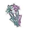
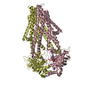
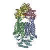
| Title | Structure of the S. cerevisiae P4B ATPase lipid flippase in the E1-ATP state | ||||||
 Components Components | Probable phospholipid-transporting ATPase NEO1 | ||||||
 Keywords Keywords |  TRANSLOCASE / P4B ATPase lipid flippase TRANSLOCASE / P4B ATPase lipid flippase | ||||||
| Function / homology |  Function and homology information Function and homology informationlysophosphatidylserine flippase activity / trans-Golgi network membrane organization / Ion transport by P-type ATPases / phosphatidylserine flippase activity / ATPase-coupled intramembrane lipid transporter activity / phosphatidylserine floppase activity / vacuole organization / phosphatidylethanolamine flippase activity / P-type phospholipid transporter / retrograde vesicle-mediated transport, Golgi to endoplasmic reticulum ...lysophosphatidylserine flippase activity / trans-Golgi network membrane organization / Ion transport by P-type ATPases / phosphatidylserine flippase activity / ATPase-coupled intramembrane lipid transporter activity / phosphatidylserine floppase activity / vacuole organization / phosphatidylethanolamine flippase activity / P-type phospholipid transporter / retrograde vesicle-mediated transport, Golgi to endoplasmic reticulum / phospholipid translocation / trans-Golgi network / endocytosis / late endosome / protein transport / endosome membrane / endosome / Golgi membrane / Golgi apparatus / magnesium ion binding / ATP hydrolysis activity / ATP binding / plasma membraneSimilarity search - Function | ||||||
| Biological species |  Saccharomyces cerevisiae (brewer's yeast) Saccharomyces cerevisiae (brewer's yeast) | ||||||
| Method | ELECTRON MICROSCOPY / single particle reconstruction / cryo EM / Resolution: 5.64 Å | ||||||
 Authors Authors | Bai, L. / Jain, B.K. / You, Q. / Duan, H.D. / Graham, T.R. / Li, H. | ||||||
| Funding support |  United States, 1items United States, 1items
| ||||||
 Citation Citation | Journal: Nat Commun / Year: 2021 Title: Structural basis of the P4B ATPase lipid flippase activity. Authors: Lin Bai / Bhawik K Jain / Qinglong You / H Diessel Duan / Mehmet Takar / Todd R Graham / Huilin Li /  Abstract: P4 ATPases are lipid flippases that are phylogenetically grouped into P4A, P4B and P4C clades. The P4A ATPases are heterodimers composed of a catalytic α-subunit and accessory β-subunit, and the ...P4 ATPases are lipid flippases that are phylogenetically grouped into P4A, P4B and P4C clades. The P4A ATPases are heterodimers composed of a catalytic α-subunit and accessory β-subunit, and the structures of several heterodimeric flippases have been reported. The S. cerevisiae Neo1 and its orthologs represent the P4B ATPases, which function as monomeric flippases without a β-subunit. It has been unclear whether monomeric flippases retain the architecture and transport mechanism of the dimeric flippases. Here we report the structure of a P4B ATPase, Neo1, in its E1-ATP, E2P-transition, and E2P states. The structure reveals a conserved architecture as well as highly similar functional intermediate states relative to dimeric flippases. Consistently, structure-guided mutagenesis of residues in the proposed substrate translocation path disrupted Neo1's ability to establish membrane asymmetry. These observations indicate that evolutionarily distant P4 ATPases use a structurally conserved mechanism for substrate transport. | ||||||
| History |
|
- Structure visualization
| Movie |
Movie viewer |
|---|---|
| Structure viewer | Molecule: MolmilJmol/JSmol |
- Downloads & links
-Download
| PDBx/mmCIF format | 7rd8.cif.gz | 181.4 KB | Display | PDBx/mmCIF format |
|---|---|---|---|---|
| PDB format | pdb7rd8.ent.gz | 138.7 KB | Display | PDB format |
| PDBx/mmJSON format | 7rd8.json.gz | Tree view | PDBx/mmJSON format | |
| Others |  Other downloads Other downloads |
-Validation report
| Arichive directory | https://data.pdbj.org/pub/pdb/validation_reports/rd/7rd8ftp://data.pdbj.org/pub/pdb/validation_reports/rd/7rd8 | HTTPS FTP |
|---|
-Related structure data
| Related structure data |  24415MC 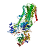 7rd6C 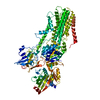 7rd7C M: map data used to model this data C: citing same article ( |
|---|---|
| Similar structure data |


-Links
 PDBj
PDBj


- Assembly
| Deposited unit | 
|
|---|---|
| 1 |
|
-Components
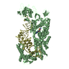
| #1: Protein | Mass: 130363.492 Da / Num. of mol.: 1 Source method: isolated from a genetically manipulated source Source: (gene. exp.) Saccharomyces cerevisiae (brewer's yeast)Gene: NEO1, YIL048W / Production host: Saccharomyces cerevisiae (brewer's yeast)References: UniProt: P40527, P-type phospholipid transporter |
|---|---|
| #2: Chemical | ChemComp-ACP /   Mass: 505.208 Da / Num. of mol.: 1 / Source method: obtained synthetically / Formula: C11H18N5O12P3 / Comment: AMP-PCP, energy-carrying molecule analogue*YM Mass: 505.208 Da / Num. of mol.: 1 / Source method: obtained synthetically / Formula: C11H18N5O12P3 / Comment: AMP-PCP, energy-carrying molecule analogue*YM |
| #3: Chemical | ChemComp-MG /   Mass: 24.305 Da / Num. of mol.: 1 / Source method: obtained synthetically / Formula: Mg Mass: 24.305 Da / Num. of mol.: 1 / Source method: obtained synthetically / Formula: Mg |
| Has ligand of interest | N |
-Experimental details
-Experiment
| Experiment | Method: ELECTRON MICROSCOPY |
|---|---|
| EM experiment | Aggregation state: PARTICLE / 3D reconstruction method: single particle reconstruction |
- Sample preparation
| Component | Name: P4B ATPase lipid flippase in the E1-ATP state / Type: COMPLEX / Entity ID: #1 / Source: RECOMBINANT |
|---|---|
| Source (natural) | Organism: Saccharomyces cerevisiae (brewer's yeast) |
| Source (recombinant) | Organism: Saccharomyces cerevisiae (brewer's yeast) |
| Buffer solution | pH: 7.4 |
| Specimen | Embedding applied: NO / Shadowing applied: NO / Staining applied: NO / Vitrification applied: YES |
| Vitrification | Cryogen name: ETHANE |
- Electron microscopy imaging
| Experimental equipment |  Model: Titan Krios / Image courtesy: FEI Company |
|---|---|
| Microscopy | Model: FEI TITAN KRIOS |
| Electron gun | Electron source: FIELD EMISSION GUN / Accelerating voltage: 300 kV / Illumination mode: FLOOD BEAM |
| Electron lens | Mode: DIFFRACTION |
| Image recording | Electron dose: 60 e/Å2 / Film or detector model: GATAN K3 (6k x 4k) |
- Processing
| Software | Name: PHENIX / Version: 1.18.2_3874: / Classification: refinement | ||||||||||||||||||||||||
|---|---|---|---|---|---|---|---|---|---|---|---|---|---|---|---|---|---|---|---|---|---|---|---|---|---|
| CTF correction | Type: NONE | ||||||||||||||||||||||||
| 3D reconstruction | Resolution: 5.64 Å / Resolution method: FSC 0.143 CUT-OFF / Num. of particles: 264891 / Symmetry type: POINT | ||||||||||||||||||||||||
| Refine LS restraints |
|
