 Movie
Movie Controller

[English] 日本語
 Yorodumi
- PDB-7r1q: X-ray structure of the adduct formed upon reaction of the gold(I)... -
+ Open data

- Basic information
| Entry | Database: PDB / ID: 7r1q | ||||||
|---|---|---|---|---|---|---|---|
| Title | X-ray structure of the adduct formed upon reaction of the gold(I) N-heterocyclic carbene complex Au2 with lysozyme | ||||||
 Components Components | Lysozyme | ||||||
 Keywords Keywords | HYDROLASE / metallodrug / protein-metal interaction / gold / carbene / anticancer | ||||||
| Function / homology |  Function and homology informationAntimicrobial peptides / Neutrophil degranulation / beta-N-acetylglucosaminidase activity / cell wall macromolecule catabolic process / lysozyme / lysozyme activity / killing of cells of another organism / defense response to Gram-negative bacterium / defense response to Gram-positive bacterium / defense response to bacterium ...Antimicrobial peptides / Neutrophil degranulation / beta-N-acetylglucosaminidase activity / cell wall macromolecule catabolic process / lysozyme / lysozyme activity / killing of cells of another organism / defense response to Gram-negative bacterium / defense response to Gram-positive bacterium / defense response to bacterium / endoplasmic reticulum / extracellular space / identical protein binding / cytoplasm Function and homology informationAntimicrobial peptides / Neutrophil degranulation / beta-N-acetylglucosaminidase activity / cell wall macromolecule catabolic process / lysozyme / lysozyme activity / killing of cells of another organism / defense response to Gram-negative bacterium / defense response to Gram-positive bacterium / defense response to bacterium ...Antimicrobial peptides / Neutrophil degranulation / beta-N-acetylglucosaminidase activity / cell wall macromolecule catabolic process / lysozyme / lysozyme activity / killing of cells of another organism / defense response to Gram-negative bacterium / defense response to Gram-positive bacterium / defense response to bacterium / endoplasmic reticulum / extracellular space / identical protein binding / cytoplasmSimilarity search - Function | ||||||
| Biological species |  Gallus gallus (chicken) Gallus gallus (chicken) | ||||||
| Method | X-RAY DIFFRACTION / SYNCHROTRON / MOLECULAR REPLACEMENT / Resolution: 1.1 Å | ||||||
 Authors Authors | Merlino, A. / Ferraro, G. | ||||||
| Funding support | 1items
| ||||||
 Citation Citation | Journal: Dalton Trans / Year: 2022 Title: Halo complexes of gold(I) containing glycoconjugate carbene ligands: synthesis, characterization, cytotoxicity and interaction with proteins and DNA model systems. Authors: Annunziata, A. / Ferraro, G. / Cucciolito, M.E. / Imbimbo, P. / Tuzi, A. / Monti, D.M. / Merlino, A. / Ruffo, F. | ||||||
| History |
|
- Structure visualization
| Structure viewer | Molecule: MolmilJmol/JSmol |
|---|
- Downloads & links
-Download
| PDBx/mmCIF format | 7r1q.cif.gz | 78.8 KB | Display | PDBx/mmCIF format |
|---|---|---|---|---|
| PDB format | pdb7r1q.ent.gz | Display | PDB format | |
| PDBx/mmJSON format | 7r1q.json.gz | Tree view | PDBx/mmJSON format | |
| Others |  Other downloads Other downloads |
-Validation report
| Arichive directory | https://data.pdbj.org/pub/pdb/validation_reports/r1/7r1qftp://data.pdbj.org/pub/pdb/validation_reports/r1/7r1q | HTTPS FTP |
|---|
-Related structure data
| Related structure data | 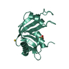 7r1pC 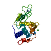 193lS S: Starting model for refinement C: citing same article ( |
|---|---|
| Similar structure data |
-Links
 PDBj
PDBj


- Assembly
| Deposited unit | 
| |||||||||||||||
|---|---|---|---|---|---|---|---|---|---|---|---|---|---|---|---|---|
| 1 |
| |||||||||||||||
| Unit cell |
| |||||||||||||||
| Components on special symmetry positions |
|
-Components
| #1: Protein | Mass: 14331.160 Da / Num. of mol.: 1 / Source method: isolated from a natural source / Source: (natural) Gallus gallus (chicken) / References: UniProt: P00698, lysozyme | ||||
|---|---|---|---|---|---|
| #2: Chemical | ChemComp-DMS / Dimethyl sulfoxide  Mass: 78.133 Da / Num. of mol.: 1 / Source method: obtained synthetically / Formula: C2H6OS / Comment: DMSO, precipitant*YM Mass: 78.133 Da / Num. of mol.: 1 / Source method: obtained synthetically / Formula: C2H6OS / Comment: DMSO, precipitant*YM | ||||
| #3: Chemical |   Mass: 196.967 Da / Num. of mol.: 3 / Source method: obtained synthetically / Formula: Au / Feature type: SUBJECT OF INVESTIGATION Mass: 196.967 Da / Num. of mol.: 3 / Source method: obtained synthetically / Formula: Au / Feature type: SUBJECT OF INVESTIGATION#4: Water | ChemComp-HOH / | Water Mass: 18.015 Da / Num. of mol.: 213 / Source method: isolated from a natural source / Formula: H2O Mass: 18.015 Da / Num. of mol.: 213 / Source method: isolated from a natural source / Formula: H2OHas ligand of interest | Y | |
-Experimental details
-Experiment
| Experiment | Method: X-RAY DIFFRACTION / Number of used crystals: 1 |
|---|
- Sample preparation
| Crystal | Density Matthews: 1.93 Å3/Da / Density % sol: 36.18 % |
|---|---|
| Crystal grow | Temperature: 293 K / Method: vapor diffusion, hanging drop / pH: 7.5 / Details: 2.0 M sodium formate 0.1 M hepes buffer pH 7.5 |
-Data collection
| Diffraction | Mean temperature: 100 K / Serial crystal experiment: N |
|---|---|
| Diffraction source | Source: SYNCHROTRON / Site: ELETTRA  / Beamline: 11.2C / Wavelength: 0.96 Å / Beamline: 11.2C / Wavelength: 0.96 Å |
| Detector | Type: DECTRIS PILATUS 6M / Detector: PIXEL / Date: Jul 27, 2021 |
| Radiation | Protocol: SINGLE WAVELENGTH / Monochromatic (M) / Laue (L): M / Scattering type: x-ray |
| Radiation wavelength | Wavelength: 0.96 Å / Relative weight: 1 |
| Reflection | Resolution: 1.1→34.43 Å / Num. obs: 44982 / % possible obs: 95.6 % / Redundancy: 17.9 % / CC1/2: 0.999 / Rmerge(I) obs: 0.059 / Rpim(I) all: 0.013 / Rrim(I) all: 0.062 / Net I/σ(I): 28.4 |
| Reflection shell | Resolution: 1.1→1.11 Å / Redundancy: 3.7 % / Rmerge(I) obs: 0.551 / Mean I/σ(I) obs: 2.4 / Num. unique obs: 1372 / CC1/2: 0.635 / Rpim(I) all: 0.318 / Rrim(I) all: 0.637 / % possible all: 58.8 |
- Processing
| Software |
| ||||||||||||||||||||||||||||||||||||||||||||||||||||||||||||||||||||||||||||||||||||||||||||||||||||||||||||||||||||||||||||||||||||||||||||||||||||||||||||||||||||||||||
|---|---|---|---|---|---|---|---|---|---|---|---|---|---|---|---|---|---|---|---|---|---|---|---|---|---|---|---|---|---|---|---|---|---|---|---|---|---|---|---|---|---|---|---|---|---|---|---|---|---|---|---|---|---|---|---|---|---|---|---|---|---|---|---|---|---|---|---|---|---|---|---|---|---|---|---|---|---|---|---|---|---|---|---|---|---|---|---|---|---|---|---|---|---|---|---|---|---|---|---|---|---|---|---|---|---|---|---|---|---|---|---|---|---|---|---|---|---|---|---|---|---|---|---|---|---|---|---|---|---|---|---|---|---|---|---|---|---|---|---|---|---|---|---|---|---|---|---|---|---|---|---|---|---|---|---|---|---|---|---|---|---|---|---|---|---|---|---|---|---|---|---|
| Refinement | Method to determine structure: MOLECULAR REPLACEMENT Starting model: 193L Resolution: 1.1→34.43 Å / Cor.coef. Fo:Fc: 0.979 / Cor.coef. Fo:Fc free: 0.965 / SU B: 0.972 / SU ML: 0.022 / Cross valid method: FREE R-VALUE / ESU R: 0.032 / ESU R Free: 0.035 Details: Hydrogens have been added in their riding positions
| ||||||||||||||||||||||||||||||||||||||||||||||||||||||||||||||||||||||||||||||||||||||||||||||||||||||||||||||||||||||||||||||||||||||||||||||||||||||||||||||||||||||||||
| Solvent computation | Ion probe radii: 0.8 Å / Shrinkage radii: 0.8 Å / VDW probe radii: 1.2 Å / Solvent model: MASK BULK SOLVENT | ||||||||||||||||||||||||||||||||||||||||||||||||||||||||||||||||||||||||||||||||||||||||||||||||||||||||||||||||||||||||||||||||||||||||||||||||||||||||||||||||||||||||||
| Displacement parameters | Biso mean: 16.618 Å2
| ||||||||||||||||||||||||||||||||||||||||||||||||||||||||||||||||||||||||||||||||||||||||||||||||||||||||||||||||||||||||||||||||||||||||||||||||||||||||||||||||||||||||||
| Refinement step | Cycle: LAST / Resolution: 1.1→34.43 Å
| ||||||||||||||||||||||||||||||||||||||||||||||||||||||||||||||||||||||||||||||||||||||||||||||||||||||||||||||||||||||||||||||||||||||||||||||||||||||||||||||||||||||||||
| Refine LS restraints |
| ||||||||||||||||||||||||||||||||||||||||||||||||||||||||||||||||||||||||||||||||||||||||||||||||||||||||||||||||||||||||||||||||||||||||||||||||||||||||||||||||||||||||||
| LS refinement shell |
|
