 Movie
Movie Controller

[English] 日本語
 Yorodumi
- PDB-7qzp: Identification and characterization of an RRM-containing, ELAV-li... -
+ Open data

- Basic information
| Entry | Database: PDB / ID: 7qzp | ||||||
|---|---|---|---|---|---|---|---|
| Title | Identification and characterization of an RRM-containing, ELAV-like, RNA binding protein in Acinetobacter Baumannii | ||||||
 Components Components | Hypothetical RNA binding protein from Acinetobacter baumannii | ||||||
 Keywords Keywords |  RNA BINDING PROTEIN / RNA / Acinetobacter / RRM / HUR RNA BINDING PROTEIN / RNA / Acinetobacter / RRM / HUR | ||||||
| Function / homology | RNA recognition motif / RNA recognition motif / Eukaryotic RNA Recognition Motif (RRM) profile. / RNA recognition motif domain / RNA-binding domain superfamily / Nucleotide-binding alpha-beta plait domain superfamily / RNA binding / RRM domain-containing protein Function and homology information Function and homology information | ||||||
| Biological species |  Acinetobacter baumannii (bacteria) Acinetobacter baumannii (bacteria) | ||||||
| Method | X-RAY DIFFRACTION / MOLECULAR REPLACEMENT / Resolution: 1.65 Å | ||||||
 Authors Authors | Ciani, C. / Perez-Rafols, A. / Bonomo, I. / Micaelli, M. / Esposito, A. / Zucal, C. / Belli, R. / D'Agostino, V.G. / Bianconi, I. / Calderone, V. ...Ciani, C. / Perez-Rafols, A. / Bonomo, I. / Micaelli, M. / Esposito, A. / Zucal, C. / Belli, R. / D'Agostino, V.G. / Bianconi, I. / Calderone, V. / Cerofolini, L. / Fragai, M. / Provenzani, A. | ||||||
| Funding support | European Union, 1items
| ||||||
 Citation Citation | Journal: Biomolecules / Year: 2022 Title: Identification and Characterization of an RRM-Containing, RNA Binding Protein in Acinetobacter baumannii . Authors: Ciani, C. / Perez-Rafols, A. / Bonomo, I. / Micaelli, M. / Esposito, A. / Zucal, C. / Belli, R. / D'Agostino, V.G. / Bianconi, I. / Calderone, V. / Cerofolini, L. / Massidda, O. / Whalen, M. ...Authors: Ciani, C. / Perez-Rafols, A. / Bonomo, I. / Micaelli, M. / Esposito, A. / Zucal, C. / Belli, R. / D'Agostino, V.G. / Bianconi, I. / Calderone, V. / Cerofolini, L. / Massidda, O. / Whalen, M.B. / Fragai, M. / Provenzani, A. | ||||||
| History |
|
- Structure visualization
| Structure viewer | Molecule: MolmilJmol/JSmol |
|---|
- Downloads & links
-Download
| PDBx/mmCIF format | 7qzp.cif.gz | 47 KB | Display | PDBx/mmCIF format |
|---|---|---|---|---|
| PDB format | pdb7qzp.ent.gz | 31.7 KB | Display | PDB format |
| PDBx/mmJSON format | 7qzp.json.gz | Tree view | PDBx/mmJSON format | |
| Others |  Other downloads Other downloads |
-Validation report
| Arichive directory | https://data.pdbj.org/pub/pdb/validation_reports/qz/7qzpftp://data.pdbj.org/pub/pdb/validation_reports/qz/7qzp | HTTPS FTP |
|---|
-Related structure data
| Related structure data | 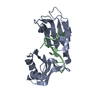 1fxlS S: Starting model for refinement |
|---|---|
| Similar structure data |
-Links
 PDBj
PDBj- Assembly
| Deposited unit | 
| ||||||||
|---|---|---|---|---|---|---|---|---|---|
| 1 |
| ||||||||
| Unit cell |
|
-Components
| #1: Protein | Mass: 10975.097 Da / Num. of mol.: 1 Source method: isolated from a genetically manipulated source Source: (gene. exp.) Acinetobacter baumannii (bacteria) / Gene: HMPREF0022_00746 / Production host: Escherichia coli (E. coli) / References: UniProt: A0A828SWT5 |
|---|---|
| #2: Water | ChemComp-HOH / Water Mass: 18.015 Da / Num. of mol.: 45 / Source method: isolated from a natural source / Formula: H2O Mass: 18.015 Da / Num. of mol.: 45 / Source method: isolated from a natural source / Formula: H2O |
-Experimental details
-Experiment
| Experiment | Method: X-RAY DIFFRACTION / Number of used crystals: 1 |
|---|
- Sample preparation
| Crystal | Density Matthews: 1.79 Å3/Da / Density % sol: 31.4 % |
|---|---|
| Crystal grow | Temperature: 293 K / Method: vapor diffusion, sitting drop / pH: 4.5 Details: 0.1 M sodium acetate trihydrate, 3 M sodium chloride |
-Data collection
| Diffraction | Mean temperature: 100 K / Serial crystal experiment: N |
|---|---|
| Diffraction source | Source: SEALED TUBE / Type: BRUKER D8 QUEST / Wavelength: 1.5418 Å |
| Detector | Type: Bruker PHOTON III / Detector: PIXEL / Date: Mar 2, 2021 |
| Radiation | Protocol: SINGLE WAVELENGTH / Monochromatic (M) / Laue (L): M / Scattering type: x-ray |
| Radiation wavelength | Wavelength: 1.5418 Å / Relative weight: 1 |
| Reflection | Resolution: 1.65→19 Å / Num. obs: 8220 / % possible obs: 87.1 % / Observed criterion σ(F): 0 / Observed criterion σ(I): 0 / Redundancy: 11.7 % / CC1/2: 0.99 / Rsym value: 0.079 / Net I/σ(I): 18.76 |
| Reflection shell | Resolution: 1.65→1.75 Å / Mean I/σ(I) obs: 1.7 / Num. unique obs: 857 / CC1/2: 0.53 / Rsym value: 0.85 |
- Processing
| Software |
| ||||||||||||||||||||||||||||||||||||||||
|---|---|---|---|---|---|---|---|---|---|---|---|---|---|---|---|---|---|---|---|---|---|---|---|---|---|---|---|---|---|---|---|---|---|---|---|---|---|---|---|---|---|
| Refinement | Method to determine structure: MOLECULAR REPLACEMENT Starting model: 1fxl Resolution: 1.65→18.87 Å / SU ML: 0.24 / Cross valid method: THROUGHOUT / σ(F): 1.36 / Phase error: 31.38 / Stereochemistry target values: ML
| ||||||||||||||||||||||||||||||||||||||||
| Solvent computation | Shrinkage radii: 0.9 Å / VDW probe radii: 1.11 Å / Solvent model: FLAT BULK SOLVENT MODEL | ||||||||||||||||||||||||||||||||||||||||
| Displacement parameters | Biso max: 106.02 Å2 / Biso mean: 36.745 Å2 / Biso min: 16.47 Å2 | ||||||||||||||||||||||||||||||||||||||||
| Refinement step | Cycle: final / Resolution: 1.65→18.87 Å
| ||||||||||||||||||||||||||||||||||||||||
| LS refinement shell | Refine-ID: X-RAY DIFFRACTION / Rfactor Rfree error: 0 / Total num. of bins used: 3
| ||||||||||||||||||||||||||||||||||||||||
| Refinement TLS params. | Method: refined / Origin x: 31.3397 Å / Origin y: 25.2325 Å / Origin z: -7.0562 Å
| ||||||||||||||||||||||||||||||||||||||||
| Refinement TLS group |
|
