 Movie
Movie Controller

[English] 日本語
 Yorodumi
- PDB-7qot: Factor XI and Plasma Kallikrein apple domain structures reveals d... -
+ Open data

- Basic information
| Entry | Database: PDB / ID: 7qot | ||||||
|---|---|---|---|---|---|---|---|
| Title | Factor XI and Plasma Kallikrein apple domain structures reveals different kininogen bound complexes | ||||||
 Components Components |
| ||||||
 Keywords Keywords |  BLOOD CLOTTING / Plasma kallikrein (PK) / High molecular weight kininogen (HK) / Factor XII (FXII) / Factor XI (FXI) / Factor IX (FIX) / bradykinin (BK) BLOOD CLOTTING / Plasma kallikrein (PK) / High molecular weight kininogen (HK) / Factor XII (FXII) / Factor XI (FXI) / Factor IX (FIX) / bradykinin (BK) | ||||||
| Function / homology |  Function and homology informationcoagulation factor XIa / serine-type aminopeptidase activity / Defective F9 activation / positive regulation of fibrinolysis / negative regulation of cell adhesion / negative regulation of blood coagulation / cysteine-type endopeptidase inhibitor activity / plasminogen activation / Intrinsic Pathway of Fibrin Clot Formation / Peptide ligand-binding receptors ...coagulation factor XIa / serine-type aminopeptidase activity / Defective F9 activation / positive regulation of fibrinolysis / negative regulation of cell adhesion / negative regulation of blood coagulation / cysteine-type endopeptidase inhibitor activity / plasminogen activation / Intrinsic Pathway of Fibrin Clot Formation / Peptide ligand-binding receptors / platelet alpha granule lumen / negative regulation of proteolysis / Post-translational protein phosphorylation / hormone activity / vasodilation / Regulation of Insulin-like Growth Factor (IGF) transport and uptake by Insulin-like Growth Factor Binding Proteins (IGFBPs) / blood coagulation / Platelet degranulation / heparin binding / G alpha (i) signalling events / positive regulation of cytosolic calcium ion concentration / G alpha (q) signalling events / collagen-containing extracellular matrix / blood microparticle / inflammatory response / positive regulation of apoptotic process / endoplasmic reticulum lumen / signaling receptor binding / serine-type endopeptidase activity / extracellular space / extracellular exosome / zinc ion binding / extracellular region / membrane / identical protein binding / plasma membrane Function and homology informationcoagulation factor XIa / serine-type aminopeptidase activity / Defective F9 activation / positive regulation of fibrinolysis / negative regulation of cell adhesion / negative regulation of blood coagulation / cysteine-type endopeptidase inhibitor activity / plasminogen activation / Intrinsic Pathway of Fibrin Clot Formation / Peptide ligand-binding receptors ...coagulation factor XIa / serine-type aminopeptidase activity / Defective F9 activation / positive regulation of fibrinolysis / negative regulation of cell adhesion / negative regulation of blood coagulation / cysteine-type endopeptidase inhibitor activity / plasminogen activation / Intrinsic Pathway of Fibrin Clot Formation / Peptide ligand-binding receptors / platelet alpha granule lumen / negative regulation of proteolysis / Post-translational protein phosphorylation / hormone activity / vasodilation / Regulation of Insulin-like Growth Factor (IGF) transport and uptake by Insulin-like Growth Factor Binding Proteins (IGFBPs) / blood coagulation / Platelet degranulation / heparin binding / G alpha (i) signalling events / positive regulation of cytosolic calcium ion concentration / G alpha (q) signalling events / collagen-containing extracellular matrix / blood microparticle / inflammatory response / positive regulation of apoptotic process / endoplasmic reticulum lumen / signaling receptor binding / serine-type endopeptidase activity / extracellular space / extracellular exosome / zinc ion binding / extracellular region / membrane / identical protein binding / plasma membraneSimilarity search - Function | ||||||
| Biological species |  Homo sapiens (human) Homo sapiens (human) | ||||||
| Method | X-RAY DIFFRACTION / SYNCHROTRON / MOLECULAR REPLACEMENT / molecular replacement / Resolution: 3.24 Å | ||||||
 Authors Authors | Li, C. / Awital, B. / Wong, S. / Dreveny, I. / Meijers, J. / Emsley, J. | ||||||
| Funding support |  United Kingdom, 1items United Kingdom, 1items
| ||||||
 Citation Citation | Journal: J Thromb Haemost / Year: 2019 Title: Plasma kallikrein structure reveals apple domain disc rotated conformation compared to factor XI. Authors: Li, C. / Voos, K.M. / Pathak, M. / Hall, G. / McCrae, K.R. / Dreveny, I. / Li, R. / Emsley, J. | ||||||
| History |
|
- Structure visualization
| Structure viewer | Molecule: MolmilJmol/JSmol |
|---|
- Downloads & links
-Download
| PDBx/mmCIF format | 7qot.cif.gz | 160.2 KB | Display | PDBx/mmCIF format |
|---|---|---|---|---|
| PDB format | pdb7qot.ent.gz | 125.6 KB | Display | PDB format |
| PDBx/mmJSON format | 7qot.json.gz | Tree view | PDBx/mmJSON format | |
| Others |  Other downloads Other downloads |
-Validation report
| Arichive directory | https://data.pdbj.org/pub/pdb/validation_reports/qo/7qotftp://data.pdbj.org/pub/pdb/validation_reports/qo/7qot | HTTPS FTP |
|---|
-Related structure data
| Related structure data |  6i44C 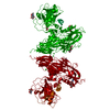 6i58C 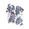 7qoxC  2f83  4bdxS S: Starting model for refinement C: citing same article ( |
|---|---|
| Similar structure data |
-Links
 PDBj
PDBj


- Assembly
| Deposited unit | 
| |||||||||||||||||||||||||||||||||||||||||||||||||||||||||||||||||||||||
|---|---|---|---|---|---|---|---|---|---|---|---|---|---|---|---|---|---|---|---|---|---|---|---|---|---|---|---|---|---|---|---|---|---|---|---|---|---|---|---|---|---|---|---|---|---|---|---|---|---|---|---|---|---|---|---|---|---|---|---|---|---|---|---|---|---|---|---|---|---|---|---|---|
| 1 | 
| |||||||||||||||||||||||||||||||||||||||||||||||||||||||||||||||||||||||
| 2 | 
| |||||||||||||||||||||||||||||||||||||||||||||||||||||||||||||||||||||||
| Unit cell |
| |||||||||||||||||||||||||||||||||||||||||||||||||||||||||||||||||||||||
| Noncrystallographic symmetry (NCS) | NCS domain:
NCS domain segments: Ens-ID: 1
|