 Movie
Movie Controller

[English] 日本語
 Yorodumi
- PDB-7qgt: Crystal structure of human cystathionine beta-synthase (delta516-... -
+ Open data

- Basic information
| Entry | Database: PDB / ID: 7qgt | ||||||
|---|---|---|---|---|---|---|---|
| Title | Crystal structure of human cystathionine beta-synthase (delta516-525) in complex with AOAA. | ||||||
 Components Components | Cystathionine beta-synthase Cystathionine beta synthase Cystathionine beta synthase | ||||||
 Keywords Keywords | LYASE / METHIONINE CYCLE / METABOLIC PATHWAY / SERINE METABOLISM | ||||||
| Function / homology |  Function and homology information Function and homology informationCysteine formation from homocysteine / homocysteine catabolic process / modified amino acid binding / cystathionine beta-synthase / cysteine biosynthetic process via cystathionine / cystathionine beta-synthase activity / Metabolism of ingested SeMet, Sec, MeSec into H2Se / homocysteine metabolic process / carbon monoxide binding / hydrogen sulfide biosynthetic process ...Cysteine formation from homocysteine / homocysteine catabolic process / modified amino acid binding / cystathionine beta-synthase / cysteine biosynthetic process via cystathionine / cystathionine beta-synthase activity / Metabolism of ingested SeMet, Sec, MeSec into H2Se / homocysteine metabolic process / carbon monoxide binding / hydrogen sulfide biosynthetic process / L-serine catabolic process / L-serine metabolic process / cartilage development involved in endochondral bone morphogenesis / regulation of nitric oxide mediated signal transduction / cysteine biosynthetic process / L-cysteine catabolic process / cerebellum morphogenesis / nitric oxide binding / cysteine biosynthetic process from serine / DNA protection / transsulfuration / endochondral ossification / S-adenosyl-L-methionine binding / response to folic acid / nitrite reductase (NO-forming) activity / superoxide metabolic process / maternal process involved in female pregnancy / blood vessel remodeling / blood vessel diameter maintenance / oxygen binding / pyridoxal phosphate binding / cellular response to hypoxia / ubiquitin protein ligase binding / heme binding / negative regulation of apoptotic process / enzyme binding / protein homodimerization activity / identical protein binding / metal ion binding / nucleus / cytosol / cytoplasmSimilarity search - Function | ||||||
| Biological species |  Homo sapiens (human) Homo sapiens (human) | ||||||
| Method | X-RAY DIFFRACTION / SYNCHROTRON / MOLECULAR REPLACEMENT / Resolution: 2.691 Å | ||||||
 Authors Authors | Hutchin, A. / Kopec, J. / Majtan, T. / Zuhra, K. / Szabo, C. | ||||||
| Funding support | 1items
| ||||||
 Citation Citation | Journal: Cell.Mol.Life Sci. / Year: 2022 Title: H 2 S biogenesis by cystathionine beta-synthase: mechanism of inhibition by aminooxyacetic acid and unexpected role of serine. Authors: Petrosino, M. / Zuhra, K. / Kopec, J. / Hutchin, A. / Szabo, C. / Majtan, T. | ||||||
| History |
|
- Structure visualization
| Structure viewer | Molecule: MolmilJmol/JSmol |
|---|
- Downloads & links
-Download
| PDBx/mmCIF format | 7qgt.cif.gz | 409.2 KB | Display | PDBx/mmCIF format |
|---|---|---|---|---|
| PDB format | pdb7qgt.ent.gz | 332.5 KB | Display | PDB format |
| PDBx/mmJSON format | 7qgt.json.gz | Tree view | PDBx/mmJSON format | |
| Others |  Other downloads Other downloads |
-Validation report
| Arichive directory | https://data.pdbj.org/pub/pdb/validation_reports/qg/7qgtftp://data.pdbj.org/pub/pdb/validation_reports/qg/7qgt | HTTPS FTP |
|---|
-Related structure data
| Related structure data | 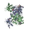 4cooS S: Starting model for refinement |
|---|---|
| Similar structure data |
-Links
 PDBj
PDBj

- Assembly
| Deposited unit | 
| ||||||||
|---|---|---|---|---|---|---|---|---|---|
| 1 |
| ||||||||
| Unit cell |
|
-Components
| #1: Protein | Cystathionine beta synthase / Beta-thionase / Serine sulfhydrase Mass: 62132.949 Da / Num. of mol.: 2 Source method: isolated from a genetically manipulated source Source: (gene. exp.) Homo sapiens (human) / Gene: CBS / Production host:  Escherichia coli BL21(DE3) (bacteria) / References: UniProt: P35520, cystathionine beta-synthase Escherichia coli BL21(DE3) (bacteria) / References: UniProt: P35520, cystathionine beta-synthase#2: Chemical | Heme B  Mass: 616.487 Da / Num. of mol.: 2 / Source method: obtained synthetically / Formula: C34H32FeN4O4 / Feature type: SUBJECT OF INVESTIGATION Mass: 616.487 Da / Num. of mol.: 2 / Source method: obtained synthetically / Formula: C34H32FeN4O4 / Feature type: SUBJECT OF INVESTIGATION#3: Chemical | ChemComp-PLP / | Pyridoxal phosphate  Mass: 247.142 Da / Num. of mol.: 1 / Source method: obtained synthetically / Formula: C8H10NO6P / Feature type: SUBJECT OF INVESTIGATION Mass: 247.142 Da / Num. of mol.: 1 / Source method: obtained synthetically / Formula: C8H10NO6P / Feature type: SUBJECT OF INVESTIGATION#4: Chemical | ChemComp-IK2 / |   Mass: 322.208 Da / Num. of mol.: 1 / Source method: isolated from a natural source / Formula: C10H15N2O8P / Feature type: SUBJECT OF INVESTIGATION Mass: 322.208 Da / Num. of mol.: 1 / Source method: isolated from a natural source / Formula: C10H15N2O8P / Feature type: SUBJECT OF INVESTIGATION#5: Water | ChemComp-HOH / | Water Mass: 18.015 Da / Num. of mol.: 167 / Source method: isolated from a natural source / Formula: H2O Mass: 18.015 Da / Num. of mol.: 167 / Source method: isolated from a natural source / Formula: H2OHas ligand of interest | Y | |
|---|
-Experimental details
-Experiment
| Experiment | Method: X-RAY DIFFRACTION / Number of used crystals: 1 |
|---|
- Sample preparation
| Crystal | Density Matthews: 2.88 Å3/Da / Density % sol: 57.25 % |
|---|---|
| Crystal grow | Temperature: 277 K / Method: vapor diffusion, sitting drop / Details: 140 mM Na FORMATE, 20% PEG3350 |
-Data collection
| Diffraction | Mean temperature: 100 K / Serial crystal experiment: N |
|---|---|
| Diffraction source | Source: SYNCHROTRON / Site: Diamond  / Beamline: I03 / Wavelength: 0.976284 Å / Beamline: I03 / Wavelength: 0.976284 Å |
| Detector | Type: DECTRIS EIGER X 16M / Detector: PIXEL / Date: Jul 24, 2021 |
| Radiation | Protocol: SINGLE WAVELENGTH / Monochromatic (M) / Laue (L): M / Scattering type: x-ray |
| Radiation wavelength | Wavelength: 0.976284 Å / Relative weight: 1 |
| Reflection | Resolution: 2.691→100.846 Å / Num. obs: 22433 / % possible obs: 92.9 % / Redundancy: 11.6 % / CC1/2: 0.988 / Net I/σ(I): 6.5 |
| Reflection shell | Resolution: 2.691→2.922 Å / Num. unique obs: 1123 / CC1/2: 0.668 |
- Processing
| Software |
| |||||||||||||||||||||||||||||||||||||||||||||||||||||||||||||||||||||||||||
|---|---|---|---|---|---|---|---|---|---|---|---|---|---|---|---|---|---|---|---|---|---|---|---|---|---|---|---|---|---|---|---|---|---|---|---|---|---|---|---|---|---|---|---|---|---|---|---|---|---|---|---|---|---|---|---|---|---|---|---|---|---|---|---|---|---|---|---|---|---|---|---|---|---|---|---|---|
| Refinement | Method to determine structure: MOLECULAR REPLACEMENT Starting model: 4coo Resolution: 2.691→55.97 Å / Cor.coef. Fo:Fc: 0.901 / Cor.coef. Fo:Fc free: 0.855 / Cross valid method: THROUGHOUT / SU Rfree Blow DPI: 0.463
| |||||||||||||||||||||||||||||||||||||||||||||||||||||||||||||||||||||||||||
| Displacement parameters | Biso mean: 51.78 Å2
| |||||||||||||||||||||||||||||||||||||||||||||||||||||||||||||||||||||||||||
| Refine analyze | Luzzati coordinate error obs: 0.39 Å | |||||||||||||||||||||||||||||||||||||||||||||||||||||||||||||||||||||||||||
| Refinement step | Cycle: LAST / Resolution: 2.691→55.97 Å
| |||||||||||||||||||||||||||||||||||||||||||||||||||||||||||||||||||||||||||
| Refine LS restraints |
| |||||||||||||||||||||||||||||||||||||||||||||||||||||||||||||||||||||||||||
| LS refinement shell | Resolution: 2.691→2.84 Å
| |||||||||||||||||||||||||||||||||||||||||||||||||||||||||||||||||||||||||||
| Refinement TLS params. | Refine-ID: X-RAY DIFFRACTION
| |||||||||||||||||||||||||||||||||||||||||||||||||||||||||||||||||||||||||||
| Refinement TLS group |
|
