 Movie
Movie Controller

[English] 日本語
 Yorodumi
- PDB-7qgq: Extended H/L (SLPH/SLPL) complex from C. difficile (CD630 strain)... -
+ Open data

- Basic information
| Entry | Database: PDB / ID: 7qgq | ||||||
|---|---|---|---|---|---|---|---|
| Title | Extended H/L (SLPH/SLPL) complex from C. difficile (CD630 strain) fit into R20291 S-layer negative stain map | ||||||
 Components Components | (Precursor of the S-layer proteins) x 2 | ||||||
 Keywords Keywords |  STRUCTURAL PROTEIN / Bacterial surface / S-layer STRUCTURAL PROTEIN / Bacterial surface / S-layer | ||||||
| Function / homology | Low molecular weight S layer protein, N-terminal / Low molecular weight S layer protein, N-terminal, subdomain / Low molecular weight S layer protein N terminal / Putative cell wall binding repeat 2 / ell wall binding domain 2 (CWB2) / identical protein binding / Precursor of the S-layer proteins Function and homology information Function and homology information | ||||||
| Biological species |  Clostridioides difficile 630 (bacteria) Clostridioides difficile 630 (bacteria) | ||||||
| Method | ELECTRON CRYSTALLOGRAPHY / electron crystallography / negative staining | ||||||
 Authors Authors | Banerji, O. / Wilson, J.S. / Bullough, P.A. | ||||||
| Funding support |  United Kingdom, 1items United Kingdom, 1items
| ||||||
 Citation Citation | Journal: Nat Commun / Year: 2022 Title: Structure and assembly of the S-layer in C. difficile. Authors: Paola Lanzoni-Mangutchi / Oishik Banerji / Jason Wilson / Anna Barwinska-Sendra / Joseph A Kirk / Filipa Vaz / Shauna O'Beirne / Arnaud Baslé / Kamel El Omari / Armin Wagner / Neil F ...Authors: Paola Lanzoni-Mangutchi / Oishik Banerji / Jason Wilson / Anna Barwinska-Sendra / Joseph A Kirk / Filipa Vaz / Shauna O'Beirne / Arnaud Baslé / Kamel El Omari / Armin Wagner / Neil F Fairweather / Gillian R Douce / Per A Bullough / Robert P Fagan / Paula S Salgado /  Abstract: Many bacteria and archaea possess a two-dimensional protein array, or S-layer, that covers the cell surface and plays crucial roles in cell physiology. Here, we report the crystal structure of SlpA, ...Many bacteria and archaea possess a two-dimensional protein array, or S-layer, that covers the cell surface and plays crucial roles in cell physiology. Here, we report the crystal structure of SlpA, the main S-layer protein of the bacterial pathogen Clostridioides difficile, and use electron microscopy to study S-layer organisation and assembly. The SlpA crystal lattice mimics S-layer assembly in the cell, through tiling of triangular prisms above the cell wall, interlocked by distinct ridges facing the environment. Strikingly, the array is very compact, with pores of only ~10 Å in diameter, compared to other S-layers (30-100 Å). The surface-exposed flexible ridges are partially dispensable for overall structure and assembly, although a mutant lacking this region becomes susceptible to lysozyme, an important molecule in host defence. Thus, our work gives insights into S-layer organisation and provides a basis for development of C. difficile-specific therapeutics. | ||||||
| History |
|
- Structure visualization
| Structure viewer | Molecule: MolmilJmol/JSmol |
|---|
- Downloads & links
-Download
| PDBx/mmCIF format | 7qgq.cif.gz | 2.6 MB | Display | PDBx/mmCIF format |
|---|---|---|---|---|
| PDB format | pdb7qgq.ent.gz | Display | PDB format | |
| PDBx/mmJSON format | 7qgq.json.gz | Tree view | PDBx/mmJSON format | |
| Others |  Other downloads Other downloads |
-Validation report
| Arichive directory | https://data.pdbj.org/pub/pdb/validation_reports/qg/7qgqftp://data.pdbj.org/pub/pdb/validation_reports/qg/7qgq | HTTPS FTP |
|---|
-Related structure data
| Related structure data | 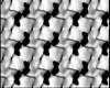 13957MC 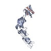 7acvC 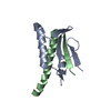 7acwC 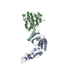 7acxC 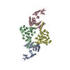 7acyC  7aczC M: map data used to model this data C: citing same article ( |
|---|---|
| Similar structure data |
-Links
 PDBj
PDBj- Assembly
| Deposited unit | 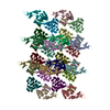
| ||||||||
|---|---|---|---|---|---|---|---|---|---|
| 1 |
| ||||||||
| Unit cell |
|
-Components
| #1: Protein | Mass: 39495.035 Da / Num. of mol.: 12 / Source method: isolated from a natural source / Source: (natural) Clostridioides difficile 630 (bacteria) / Plasmid details: RT012, SLCT7 / Strain: 630 / References: UniProt: Q183M8#2: Protein | Mass: 33949.785 Da / Num. of mol.: 12 / Source method: isolated from a natural source / Source: (natural) Clostridioides difficile 630 (bacteria) / Plasmid details: RT012, SLCT7 / Strain: 630 / References: UniProt: Q183M8 |
|---|
-Experimental details
-Experiment
| Experiment | Method: ELECTRON CRYSTALLOGRAPHY |
|---|---|
| EM experiment | Aggregation state: 2D ARRAY / 3D reconstruction method: electron crystallography |
- Sample preparation
| Component | Name: Electron crystallographic reconstruction of R20291 S-layer, extended to cover 12 molecules of SlpA for flexible fitting of X-ray structure to map Type: COMPLEX / Entity ID: all / Source: NATURAL |
|---|---|
| Molecular weight | Experimental value: NO |
| Source (natural) | Organism: Clostridioides difficile R20291 (bacteria) |
| Buffer solution | pH: 7.4 |
| Specimen | Embedding applied: NO / Shadowing applied: NO / Staining applied: YES / Vitrification applied: NO |
| EM staining | Type: NEGATIVE Details: Negatively stained EM samples were prepared by depositing sample on continuous carbon layer and staining with 2 % Uranyl Formate with blotting Material: Uranyl Formate |
| Specimen support | Grid material: COPPER / Grid mesh size: 300 divisions/in. / Grid type: Homemade |
-Data collection
| Microscopy | Model: FEI/PHILIPS CM200FEG |
|---|---|
| Electron gun | Electron source: FIELD EMISSION GUN / Accelerating voltage: 200 kV / Illumination mode: FLOOD BEAM |
| Electron lens | Mode: BRIGHT FIELDBright-field microscopy / Nominal defocus max: 2200 nm / Nominal defocus min: 800 nm |
| Image recording | Electron dose: 0.1 e/Å2 / Film or detector model: GATAN ULTRASCAN 4000 (4k x 4k) / Num. of real images: 36 / Details: Images binned by 2 before processing |
| Image scans | Sampling size: 15 µm / Width: 2048 / Height: 2048 |
| EM diffraction | Camera length: 0.1 mm |
| EM diffraction shell | Resolution: 18.25→78.79 Å / Fourier space coverage: 0.1 % / Multiplicity: 0.1 / Num. of structure factors: 710 / Phase residual: 21.9 ° |
| EM diffraction stats | Fourier space coverage: 0.1 % / High resolution: 18.25 Å / Num. of intensities measured: 1085 / Num. of structure factors: 710 / Phase error: 22.3 ° / Phase residual: 21.9 ° Phase error rejection criteria: Structure factors with phase errors higher than 45 degrees were omitted from refinement Rmerge: 0.193 |
- Processing
| EM software |
| ||||||||||||||||||||||||||||||
|---|---|---|---|---|---|---|---|---|---|---|---|---|---|---|---|---|---|---|---|---|---|---|---|---|---|---|---|---|---|---|---|
| EM 3D crystal entity | ∠α: 90 ° / ∠β: 90 ° / ∠γ: 100 ° / A: 80 Å / B: 80 Å / C: 160 Å / Space group name: P112 / Space group num: 13 | ||||||||||||||||||||||||||||||
| CTF correction | Type: PHASE FLIPPING AND AMPLITUDE CORRECTION | ||||||||||||||||||||||||||||||
| 3D reconstruction | Resolution method: DIFFRACTION PATTERN/LAYERLINES / Symmetry type: 3D CRYSTAL | ||||||||||||||||||||||||||||||
| Atomic model building | Protocol: FLEXIBLE FIT / Space: REAL | ||||||||||||||||||||||||||||||
| Refinement | Cross valid method: THROUGHOUT | ||||||||||||||||||||||||||||||
| Displacement parameters | Biso max: 104.01 Å2 / Biso mean: 57.857 Å2 / Biso min: 34.65 Å2 |
