 Movie
Movie Controller

[English] 日本語
 Yorodumi
- PDB-7qgl: Human CD73 (ecto 5'-nucleotidase) in complex with MRS4602 (a 3-me... -
+ Open data

- Basic information
| Entry | Database: PDB / ID: 7qgl | ||||||
|---|---|---|---|---|---|---|---|
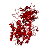
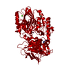
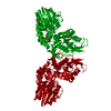
| Title | Human CD73 (ecto 5'-nucleotidase) in complex with MRS4602 (a 3-methyl-CMPCP derivative, compound 21 in paper) in the open state | ||||||
 Components Components | 5'-nucleotidase | ||||||
 Keywords Keywords | HYDROLASE / en / e5nt / ecto-nucleotidase / inhibitor | ||||||
| Function / homology |  Function and homology informationthymidylate 5'-phosphatase / thymidylate 5'-phosphatase activity / ADP catabolic process / 5'-deoxynucleotidase / 5'-deoxynucleotidase activity / 7-methylguanosine nucleotidase / inhibition of non-skeletal tissue mineralization / adenosine biosynthetic process / Pyrimidine catabolism / AMP catabolic process ...thymidylate 5'-phosphatase / thymidylate 5'-phosphatase activity / ADP catabolic process / 5'-deoxynucleotidase / 5'-deoxynucleotidase activity / 7-methylguanosine nucleotidase / inhibition of non-skeletal tissue mineralization / adenosine biosynthetic process / Pyrimidine catabolism / AMP catabolic process / GMP 5'-nucleotidase activity / IMP-specific 5'-nucleotidase / IMP 5'-nucleotidase activity / Nicotinate metabolism / Purine catabolism / XMP 5'-nucleosidase activity / 5'-nucleotidase / 5'-nucleotidase activity / leukocyte cell-cell adhesion / DNA metabolic process / response to ATP / response to inorganic substance / calcium ion homeostasis / Purinergic signaling in leishmaniasis infection / ATP metabolic process / negative regulation of inflammatory response / external side of plasma membrane / nucleotide binding / cell surface / extracellular exosome / zinc ion binding / nucleoplasm / membrane / identical protein binding / plasma membrane / cytosol Function and homology informationthymidylate 5'-phosphatase / thymidylate 5'-phosphatase activity / ADP catabolic process / 5'-deoxynucleotidase / 5'-deoxynucleotidase activity / 7-methylguanosine nucleotidase / inhibition of non-skeletal tissue mineralization / adenosine biosynthetic process / Pyrimidine catabolism / AMP catabolic process ...thymidylate 5'-phosphatase / thymidylate 5'-phosphatase activity / ADP catabolic process / 5'-deoxynucleotidase / 5'-deoxynucleotidase activity / 7-methylguanosine nucleotidase / inhibition of non-skeletal tissue mineralization / adenosine biosynthetic process / Pyrimidine catabolism / AMP catabolic process / GMP 5'-nucleotidase activity / IMP-specific 5'-nucleotidase / IMP 5'-nucleotidase activity / Nicotinate metabolism / Purine catabolism / XMP 5'-nucleosidase activity / 5'-nucleotidase / 5'-nucleotidase activity / leukocyte cell-cell adhesion / DNA metabolic process / response to ATP / response to inorganic substance / calcium ion homeostasis / Purinergic signaling in leishmaniasis infection / ATP metabolic process / negative regulation of inflammatory response / external side of plasma membrane / nucleotide binding / cell surface / extracellular exosome / zinc ion binding / nucleoplasm / membrane / identical protein binding / plasma membrane / cytosolSimilarity search - Function | ||||||
| Biological species |  Homo sapiens (human) Homo sapiens (human) | ||||||
| Method | X-RAY DIFFRACTION / FOURIER SYNTHESIS / Resolution: 1.5 Å | ||||||
 Authors Authors | Strater, N. | ||||||
| Funding support |  Germany, 1items Germany, 1items
| ||||||
 Citation Citation | Journal: J.Med.Chem. / Year: 2022 Title: Structure-Activity Relationship of 3-Methylcytidine-5'-alpha , beta-methylenediphosphates as CD73 Inhibitors. Authors: Scortichini, M. / Idris, R.M. / Moschutz, S. / Keim, A. / Salmaso, V. / Dobelmann, C. / Oliva, P. / Losenkova, K. / Irjala, H. / Vaittinen, S. / Sandholm, J. / Yegutkin, G.G. / Strater, N. / ...Authors: Scortichini, M. / Idris, R.M. / Moschutz, S. / Keim, A. / Salmaso, V. / Dobelmann, C. / Oliva, P. / Losenkova, K. / Irjala, H. / Vaittinen, S. / Sandholm, J. / Yegutkin, G.G. / Strater, N. / Junker, A. / Muller, C.E. / Jacobson, K.A. | ||||||
| History |
|
- Structure visualization
| Structure viewer | Molecule: MolmilJmol/JSmol |
|---|
- Downloads & links
-Download
| PDBx/mmCIF format | 7qgl.cif.gz | 238.7 KB | Display | PDBx/mmCIF format |
|---|---|---|---|---|
| PDB format | pdb7qgl.ent.gz | 186.1 KB | Display | PDB format |
| PDBx/mmJSON format | 7qgl.json.gz | Tree view | PDBx/mmJSON format | |
| Others |  Other downloads Other downloads |
-Validation report
| Arichive directory | https://data.pdbj.org/pub/pdb/validation_reports/qg/7qglftp://data.pdbj.org/pub/pdb/validation_reports/qg/7qgl | HTTPS FTP |
|---|
-Related structure data
| Related structure data |  7qgaC  7qgmC  7qgoC  6tveS S: Starting model for refinement C: citing same article ( |
|---|---|
| Similar structure data |


-Links
 PDBj
PDBj


- Assembly
| Deposited unit | 
| ||||||||
|---|---|---|---|---|---|---|---|---|---|
| 1 | 
| ||||||||
| Unit cell |
|
-Components
-Protein , 1 types, 1 molecules A
| #1: Protein | / 5'-NT / Ecto-5'-nucleotidase Mass: 60363.316 Da / Num. of mol.: 1 Source method: isolated from a genetically manipulated source Source: (gene. exp.) Homo sapiens (human) / Gene: NT5E, NT5, NTE / Production host: Homo sapiens (human) / References: UniProt: P21589, 5'-nucleotidase |
|---|
-Non-polymers , 5 types, 660 molecules 


| #2: Chemical |  Mass: 65.409 Da / Num. of mol.: 2 / Source method: obtained synthetically / Formula: Zn Mass: 65.409 Da / Num. of mol.: 2 / Source method: obtained synthetically / Formula: Zn#3: Chemical | ChemComp-CA / |  Mass: 40.078 Da / Num. of mol.: 1 / Source method: obtained synthetically / Formula: Ca Mass: 40.078 Da / Num. of mol.: 1 / Source method: obtained synthetically / Formula: Ca#4: Chemical | ChemComp-BW0 / [[( |  Mass: 579.388 Da / Num. of mol.: 1 / Source method: obtained synthetically / Formula: C20H27N3O13P2 / Feature type: SUBJECT OF INVESTIGATION Mass: 579.388 Da / Num. of mol.: 1 / Source method: obtained synthetically / Formula: C20H27N3O13P2 / Feature type: SUBJECT OF INVESTIGATION#5: Chemical | ChemComp-1PE / Polyethylene glycol Mass: 238.278 Da / Num. of mol.: 4 / Source method: obtained synthetically / Formula: C10H22O6 / Comment: precipitant*YM Mass: 238.278 Da / Num. of mol.: 4 / Source method: obtained synthetically / Formula: C10H22O6 / Comment: precipitant*YM#6: Water | ChemComp-HOH / | WaterMass: 18.015 Da / Num. of mol.: 652 / Source method: isolated from a natural source / Formula: H2O |
|---|
-Details
| Has ligand of interest | Y |
|---|
-Experimental details
-Experiment
| Experiment | Method: X-RAY DIFFRACTION / Number of used crystals: 1 |
|---|
- Sample preparation
| Crystal | Density Matthews: 2.37 Å3/Da / Density % sol: 48 % |
|---|---|
| Crystal grow | Temperature: 291 K / Method: vapor diffusion, hanging drop / pH: 6.2 Details: 10 mM MRS4602, 100 microM ZnCl2, 8 % PEG 6.000, 0.1 M MES pH 6.2, 20 % PEG200 |
-Data collection
| Diffraction | Mean temperature: 100 K / Serial crystal experiment: N | ||||||||||||||||||||||||||||||
|---|---|---|---|---|---|---|---|---|---|---|---|---|---|---|---|---|---|---|---|---|---|---|---|---|---|---|---|---|---|---|---|
| Diffraction source | Source: ROTATING ANODE / Type: RIGAKU MICROMAX-007 HF / Wavelength: 1.5418 Å | ||||||||||||||||||||||||||||||
| Detector | Type: RIGAKU HyPix-6000HE / Detector: PIXEL / Date: Sep 16, 2020 | ||||||||||||||||||||||||||||||
| Radiation | Protocol: SINGLE WAVELENGTH / Monochromatic (M) / Laue (L): M / Scattering type: x-ray | ||||||||||||||||||||||||||||||
| Radiation wavelength | Wavelength: 1.5418 Å / Relative weight: 1 | ||||||||||||||||||||||||||||||
| Reflection | Resolution: 1.5→21.63 Å / Num. obs: 91996 / % possible obs: 99.6 % / Redundancy: 3.9 % / Biso Wilson estimate: 12.3 Å2 / CC1/2: 0.998 / Rmerge(I) obs: 0.057 / Rpim(I) all: 0.031 / Rrim(I) all: 0.065 / Net I/σ(I): 14.8 / Num. measured all: 362347 / Scaling rejects: 217 | ||||||||||||||||||||||||||||||
| Reflection shell | Diffraction-ID: 1
|
- Processing
| Software |
| ||||||||||||||||||||||||||||||||||||||||||||||||||||||||||||||||||||||||||||||||||||||||||||||||||||||||||||
|---|---|---|---|---|---|---|---|---|---|---|---|---|---|---|---|---|---|---|---|---|---|---|---|---|---|---|---|---|---|---|---|---|---|---|---|---|---|---|---|---|---|---|---|---|---|---|---|---|---|---|---|---|---|---|---|---|---|---|---|---|---|---|---|---|---|---|---|---|---|---|---|---|---|---|---|---|---|---|---|---|---|---|---|---|---|---|---|---|---|---|---|---|---|---|---|---|---|---|---|---|---|---|---|---|---|---|---|---|---|
| Refinement | Method to determine structure: FOURIER SYNTHESIS Starting model: 6tve Resolution: 1.5→21.63 Å / Cor.coef. Fo:Fc: 0.96 / Cor.coef. Fo:Fc free: 0.956 / SU R Cruickshank DPI: 0.062 / Cross valid method: THROUGHOUT / σ(F): 0 / SU R Blow DPI: 0.067 / SU Rfree Blow DPI: 0.064 / SU Rfree Cruickshank DPI: 0.061 Details: HYDROGENS WERE FULLY REFINED WITH ZERO OCCUPANCY AT NUCLEAR POSITION. REFINEMENT NOTES. NUMBER OF REFINEMENT NOTES : 1 NOTE 1 : IDEAL-DIST CONTACT TERM CONTACT SETUP. ALL ATOMS HAVE CCP4 ATOM TYPE FROM LIBRARY
| ||||||||||||||||||||||||||||||||||||||||||||||||||||||||||||||||||||||||||||||||||||||||||||||||||||||||||||
| Displacement parameters | Biso max: 116.04 Å2 / Biso mean: 14.73 Å2 / Biso min: 4.53 Å2
| ||||||||||||||||||||||||||||||||||||||||||||||||||||||||||||||||||||||||||||||||||||||||||||||||||||||||||||
| Refine analyze | Luzzati coordinate error obs: 0.16 Å | ||||||||||||||||||||||||||||||||||||||||||||||||||||||||||||||||||||||||||||||||||||||||||||||||||||||||||||
| Refinement step | Cycle: final / Resolution: 1.5→21.63 Å
| ||||||||||||||||||||||||||||||||||||||||||||||||||||||||||||||||||||||||||||||||||||||||||||||||||||||||||||
| Refine LS restraints |
| ||||||||||||||||||||||||||||||||||||||||||||||||||||||||||||||||||||||||||||||||||||||||||||||||||||||||||||
| LS refinement shell | Resolution: 1.5→1.51 Å / Rfactor Rfree error: 0 / Total num. of bins used: 51
| ||||||||||||||||||||||||||||||||||||||||||||||||||||||||||||||||||||||||||||||||||||||||||||||||||||||||||||
| Refinement TLS params. | Method: refined / Refine-ID: X-RAY DIFFRACTION
| ||||||||||||||||||||||||||||||||||||||||||||||||||||||||||||||||||||||||||||||||||||||||||||||||||||||||||||
| Refinement TLS group |
|
