 Movie
Movie Controller

[English] 日本語
 Yorodumi
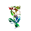
- PDB-7qeh: LTA-binding domain of SlpA, the S-layer protein from Lactobacillu... -
+ Open data

- Basic information
| Entry | Database: PDB / ID: 7qeh | ||||||
|---|---|---|---|---|---|---|---|
| Title | LTA-binding domain of SlpA, the S-layer protein from Lactobacillus amylovorus | ||||||
 Components Components | S-layer | ||||||
 Keywords Keywords | CELL ADHESION / s-layer / LTA-binding / lactobacillus amylovorus / polymer binding | ||||||
| Function / homology | Lactobacillus surface layer protein / Surface layer protein A domain / Surface layer protein A domain / structural constituent of cell wall / S-layer / peptidoglycan-based cell wall / PHOSPHATE ION / S-layer Function and homology information Function and homology information | ||||||
| Biological species |  Lactobacillus amylovorus (bacteria) Lactobacillus amylovorus (bacteria) | ||||||
| Method | X-RAY DIFFRACTION / SYNCHROTRON / SAD / Resolution: 1.67 Å | ||||||
 Authors Authors | Eder, M. / Dordic, A. / Sagmeister, T. / Pavkov-Keller, T. | ||||||
| Funding support |  Austria, 1items Austria, 1items
| ||||||
 Citation Citation | Journal: to be published Title: The binding of the lactobacilli S-layer protein to the bacterial cell through interaction with LTA Authors: Eder, M. / Gubensaek, N. / Sagmeister, T. / Grininger, C. / Vejzovic, D. / Damisch, E. / Dordic, A. / Codee, J. / Pavkov-Keller, T. | ||||||
| History |
|
- Structure visualization
| Structure viewer | Molecule: MolmilJmol/JSmol |
|---|
- Downloads & links
-Download
| PDBx/mmCIF format | 7qeh.cif.gz | 69.9 KB | Display | PDBx/mmCIF format |
|---|---|---|---|---|
| PDB format | pdb7qeh.ent.gz | 44.9 KB | Display | PDB format |
| PDBx/mmJSON format | 7qeh.json.gz | Tree view | PDBx/mmJSON format | |
| Others |  Other downloads Other downloads |
-Validation report
| Arichive directory | https://data.pdbj.org/pub/pdb/validation_reports/qe/7qehftp://data.pdbj.org/pub/pdb/validation_reports/qe/7qeh | HTTPS FTP |
|---|
-Related structure data
-Links
 PDBj
PDBj- Assembly
| Deposited unit | 
| ||||||||||||
|---|---|---|---|---|---|---|---|---|---|---|---|---|---|
| 1 |
| ||||||||||||
| Unit cell |
|
-Components
| #1: Protein | Mass: 16231.854 Da / Num. of mol.: 1 Source method: isolated from a genetically manipulated source Source: (gene. exp.) Lactobacillus amylovorus (strain GRL 1112) (bacteria)Strain: GRL 1112 / Gene: LA2_00970 / Production host: Escherichia coli (E. coli) / References: UniProt: E4SK47 |
|---|---|
| #2: Chemical | ChemComp-PO4 / Phosphate  Mass: 94.971 Da / Num. of mol.: 1 / Source method: obtained synthetically / Formula: PO4 / Feature type: SUBJECT OF INVESTIGATION Mass: 94.971 Da / Num. of mol.: 1 / Source method: obtained synthetically / Formula: PO4 / Feature type: SUBJECT OF INVESTIGATION |
| #3: Water | ChemComp-HOH / Water Mass: 18.015 Da / Num. of mol.: 75 / Source method: isolated from a natural source / Formula: H2O Mass: 18.015 Da / Num. of mol.: 75 / Source method: isolated from a natural source / Formula: H2O |
| Has ligand of interest | Y |
-Experimental details
-Experiment
| Experiment | Method: X-RAY DIFFRACTION / Number of used crystals: 1 |
|---|
- Sample preparation
| Crystal | Density Matthews: 1.83 Å3/Da / Density % sol: 32.75 % |
|---|---|
| Crystal grow | Temperature: 293 K / Method: vapor diffusion, sitting drop Details: Morpheus screen condition #2-29 (20 mM of each carboxylic acid (sodium formate, ammonium acetate, sodium citrate tribasic dehydrate, sodium potassium tartrate tetrahydrate, and sodium ...Details: Morpheus screen condition #2-29 (20 mM of each carboxylic acid (sodium formate, ammonium acetate, sodium citrate tribasic dehydrate, sodium potassium tartrate tetrahydrate, and sodium oxamate), 0.1 M sodium HEPES- MOPS pH 7.5 with 20 % v/v PEG 500 MME and 10 % w/v PEG 20000). A protein stock solution of 20 mg/mL in 10 mM HEPES pH 7 and 100 mM NaCl was used. 1uL drop with 1:1 ration protein to reservoir solution. |
-Data collection
| Diffraction | Mean temperature: 100 K / Serial crystal experiment: N |
|---|---|
| Diffraction source | Source: SYNCHROTRON / Site: ESRF  / Beamline: ID23-1 / Wavelength: 0.9789 Å / Beamline: ID23-1 / Wavelength: 0.9789 Å |
| Detector | Type: DECTRIS PILATUS 6M-F / Detector: PIXEL / Date: Mar 1, 2015 |
| Radiation | Protocol: SINGLE WAVELENGTH / Monochromatic (M) / Laue (L): M / Scattering type: x-ray |
| Radiation wavelength | Wavelength: 0.9789 Å / Relative weight: 1 |
| Reflection | Resolution: 1.67→39.08 Å / Num. obs: 26186 / % possible obs: 99.39 % / Redundancy: 12.2 % / Biso Wilson estimate: 18.71 Å2 / CC1/2: 0.999 / CC star: 1 / Rmerge(I) obs: 0.109 / Rpim(I) all: 0.032 / Rrim(I) all: 0.114 / Net I/σ(I): 19.84 |
| Reflection shell | Resolution: 1.67→1.734 Å / Rmerge(I) obs: 0.86 / Mean I/σ(I) obs: 3.2 / Num. unique obs: 1361 / CC1/2: 0.805 / CC star: 0.947 / Rpim(I) all: 0.316 |
- Processing
| Software |
| ||||||||||||||||||||||||||||||||||||||||||||||||||||||||||||||||||||||||||||||||||||||||||||||||||||||||||||||||||||||||||||||||||||||||||||
|---|---|---|---|---|---|---|---|---|---|---|---|---|---|---|---|---|---|---|---|---|---|---|---|---|---|---|---|---|---|---|---|---|---|---|---|---|---|---|---|---|---|---|---|---|---|---|---|---|---|---|---|---|---|---|---|---|---|---|---|---|---|---|---|---|---|---|---|---|---|---|---|---|---|---|---|---|---|---|---|---|---|---|---|---|---|---|---|---|---|---|---|---|---|---|---|---|---|---|---|---|---|---|---|---|---|---|---|---|---|---|---|---|---|---|---|---|---|---|---|---|---|---|---|---|---|---|---|---|---|---|---|---|---|---|---|---|---|---|---|---|---|
| Refinement | Method to determine structure: SAD / Resolution: 1.67→39.08 Å / SU ML: 0.1598 / Cross valid method: FREE R-VALUE / σ(F): 1.34 / Phase error: 17.8422 Stereochemistry target values: GeoStd + Monomer Library + CDL v1.2
| ||||||||||||||||||||||||||||||||||||||||||||||||||||||||||||||||||||||||||||||||||||||||||||||||||||||||||||||||||||||||||||||||||||||||||||
| Solvent computation | Shrinkage radii: 0.9 Å / VDW probe radii: 1.11 Å / Solvent model: FLAT BULK SOLVENT MODEL | ||||||||||||||||||||||||||||||||||||||||||||||||||||||||||||||||||||||||||||||||||||||||||||||||||||||||||||||||||||||||||||||||||||||||||||
| Displacement parameters | Biso mean: 24.47 Å2 | ||||||||||||||||||||||||||||||||||||||||||||||||||||||||||||||||||||||||||||||||||||||||||||||||||||||||||||||||||||||||||||||||||||||||||||
| Refinement step | Cycle: LAST / Resolution: 1.67→39.08 Å
| ||||||||||||||||||||||||||||||||||||||||||||||||||||||||||||||||||||||||||||||||||||||||||||||||||||||||||||||||||||||||||||||||||||||||||||
| Refine LS restraints |
| ||||||||||||||||||||||||||||||||||||||||||||||||||||||||||||||||||||||||||||||||||||||||||||||||||||||||||||||||||||||||||||||||||||||||||||
| LS refinement shell |
|
