Movie
Movie Controller

[English] 日本語
 Yorodumi
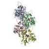
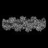
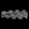
- PDB-7q8s: Leishmania major ADP-actin filament decorated with Leishmania maj... -
+ Open data

- Basic information
| Entry | Database: PDB / ID: 7q8s | |||||||||
|---|---|---|---|---|---|---|---|---|---|---|
| Title | Leishmania major ADP-actin filament decorated with Leishmania major cofilin | |||||||||
 Components Components |
| |||||||||
 Keywords Keywords |  STRUCTURAL PROTEIN / Actin / Filament / Parasite / cofilin / ADP / decorated STRUCTURAL PROTEIN / Actin / Filament / Parasite / cofilin / ADP / decorated | |||||||||
| Function / homology |  Function and homology informationkinetoplast / actin filament depolymerization / nuclear lumen / ciliary plasm / intracellular transport / actin filament binding / actin cytoskeleton / endonuclease activity / cytoskeleton / chromatin remodeling ...kinetoplast / actin filament depolymerization / nuclear lumen / ciliary plasm / intracellular transport / actin filament binding / actin cytoskeleton / endonuclease activity / cytoskeleton / chromatin remodeling / cell division / cytoplasm Function and homology informationkinetoplast / actin filament depolymerization / nuclear lumen / ciliary plasm / intracellular transport / actin filament binding / actin cytoskeleton / endonuclease activity / cytoskeleton / chromatin remodeling ...kinetoplast / actin filament depolymerization / nuclear lumen / ciliary plasm / intracellular transport / actin filament binding / actin cytoskeleton / endonuclease activity / cytoskeleton / chromatin remodeling / cell division / cytoplasmSimilarity search - Function | |||||||||
| Biological species |  Leishmania major (eukaryote) Leishmania major (eukaryote) | |||||||||
| Method | ELECTRON MICROSCOPY / helical reconstruction / cryo EM / Resolution: 3.4 Å | |||||||||
 Authors Authors | Kotila, T. / Muniyandi, S. / Lappalainen, P. / Huiskonen, J.T. | |||||||||
| Funding support |  Finland, 2items Finland, 2items
| |||||||||
 Citation Citation | Journal: Nat Commun / Year: 2022 Title: Structural basis of rapid actin dynamics in the evolutionarily divergent Leishmania parasite. Authors: Tommi Kotila / Hugo Wioland / Muniyandi Selvaraj / Konstantin Kogan / Lina Antenucci / Antoine Jégou / Juha T Huiskonen / Guillaume Romet-Lemonne / Pekka Lappalainen /  Abstract: Actin polymerization generates forces for cellular processes throughout the eukaryotic kingdom, but our understanding of the 'ancient' actin turnover machineries is limited. We show that, ...Actin polymerization generates forces for cellular processes throughout the eukaryotic kingdom, but our understanding of the 'ancient' actin turnover machineries is limited. We show that, despite > 1 billion years of evolution, pathogenic Leishmania major parasite and mammalian actins share the same overall fold and co-polymerize with each other. Interestingly, Leishmania harbors a simple actin-regulatory machinery that lacks cofilin 'cofactors', which accelerate filament disassembly in higher eukaryotes. By applying single-filament biochemistry we discovered that, compared to mammalian proteins, Leishmania actin filaments depolymerize more rapidly from both ends, and are severed > 100-fold more efficiently by cofilin. Our high-resolution cryo-EM structures of Leishmania ADP-, ADP-Pi- and cofilin-actin filaments identify specific features at actin subunit interfaces and cofilin-actin interactions that explain the unusually rapid dynamics of parasite actin filaments. Our findings reveal how divergent parasites achieve rapid actin dynamics using a remarkably simple set of actin-binding proteins, and elucidate evolution of the actin cytoskeleton. | |||||||||
| History |
|
- Structure visualization
| Structure viewer | Molecule: MolmilJmol/JSmol |
|---|
- Downloads & links
-Download
| PDBx/mmCIF format | 7q8s.cif.gz | 422.4 KB | Display | PDBx/mmCIF format |
|---|---|---|---|---|
| PDB format | pdb7q8s.ent.gz | 355.8 KB | Display | PDB format |
| PDBx/mmJSON format | 7q8s.json.gz | Tree view | PDBx/mmJSON format | |
| Others |  Other downloads Other downloads |
-Validation report
| Arichive directory | https://data.pdbj.org/pub/pdb/validation_reports/q8/7q8sftp://data.pdbj.org/pub/pdb/validation_reports/q8/7q8s | HTTPS FTP |
|---|
-Related structure data
| Related structure data | 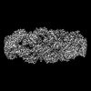 13865MC 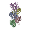 7q8bC 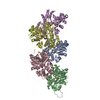 7q8cC C: citing same article ( M: map data used to model this data |
|---|---|
| Similar structure data |
-Links
 PDBj
PDBj


- Assembly
| Deposited unit | 
|
|---|---|
| 1 |
|
-Components
| #1: Protein | Mass: 42063.867 Da / Num. of mol.: 5 Source method: isolated from a genetically manipulated source Source: (gene. exp.) Leishmania major (eukaryote) / Gene: ACT, LMJF_04_1230 / Production host:   Spodoptera frugiperda (fall armyworm) / References: UniProt: Q9U1E8 Spodoptera frugiperda (fall armyworm) / References: UniProt: Q9U1E8#2: Protein | ADF/Cofilin familyMass: 15606.783 Da / Num. of mol.: 5 Source method: isolated from a genetically manipulated source Source: (gene. exp.) Leishmania major (eukaryote) / Gene: LMJF_29_0510 / Production host:  Escherichia coli BL21 (bacteria) / References: UniProt: E9ADQ2 Escherichia coli BL21 (bacteria) / References: UniProt: E9ADQ2#3: Chemical | ChemComp-ADP / Adenosine diphosphate  Mass: 427.201 Da / Num. of mol.: 5 / Source method: obtained synthetically / Formula: C10H15N5O10P2 / Feature type: SUBJECT OF INVESTIGATION / Comment: ADP, energy-carrying molecule*YM Mass: 427.201 Da / Num. of mol.: 5 / Source method: obtained synthetically / Formula: C10H15N5O10P2 / Feature type: SUBJECT OF INVESTIGATION / Comment: ADP, energy-carrying molecule*YM#4: Chemical | ChemComp-MG /   Mass: 24.305 Da / Num. of mol.: 5 / Source method: obtained synthetically / Formula: Mg / Feature type: SUBJECT OF INVESTIGATION Mass: 24.305 Da / Num. of mol.: 5 / Source method: obtained synthetically / Formula: Mg / Feature type: SUBJECT OF INVESTIGATIONHas ligand of interest | Y | |
|---|
-Experimental details
-Experiment
| Experiment | Method: ELECTRON MICROSCOPY |
|---|---|
| EM experiment | Aggregation state: FILAMENT / 3D reconstruction method: helical reconstruction |
- Sample preparation
| Component | Name: ADP-state actin filament decorated with Leishmania major cofilin. Type: COMPLEX / Entity ID: #1-#2 / Source: MULTIPLE SOURCES |
|---|---|
| Molecular weight | Units: KILODALTONS/NANOMETER / Experimental value: NO |
| Source (natural) | Organism: Leishmania major (eukaryote) |
| Source (recombinant) | Organism: Spodoptera frugiperda (fall armyworm) |
| Buffer solution | pH: 7.4 Details: 10 mM HEPES, 125 mM NaCl, 5 mM KCl, 0.2 mM ATP, 0.4 mM EGTA, 1 mM MgCl2, 1 mM DTT |
| Specimen | Embedding applied: NO / Shadowing applied: NO / Staining applied: NO / Vitrification applied: YES Details: 3 ul of 12 uM Leishmania actin was applied on the grid and left to settle for 15 s at room temperature. This grid was then mounted to the vitrobot and 1 ul of actin was withdrawn. On top of ...Details: 3 ul of 12 uM Leishmania actin was applied on the grid and left to settle for 15 s at room temperature. This grid was then mounted to the vitrobot and 1 ul of actin was withdrawn. On top of the remaining 2 ul of Leishmania actin, 1 ul of Leishmania cofilin at 77 uM |
| Specimen support | Grid material: COPPER / Grid mesh size: 300 divisions/in. / Grid type: Quantifoil R1.2/1.3 |
| Vitrification | Instrument: FEI VITROBOT MARK IV / Cryogen name: ETHANE / Humidity: 95 % / Chamber temperature: 279.15 K / Details: blot for 5 seconds before plunging, blot force 15 |
- Electron microscopy imaging
| Experimental equipment |  Model: Titan Krios / Image courtesy: FEI Company |
|---|---|
| Microscopy | Model: FEI TITAN KRIOS |
| Electron gun | Electron source: FIELD EMISSION GUN / Accelerating voltage: 300 kV / Illumination mode: FLOOD BEAM |
| Electron lens | Mode: BRIGHT FIELDBright-field microscopy |
| Image recording | Electron dose: 55 e/Å2 / Film or detector model: GATAN K3 BIOQUANTUM (6k x 4k) / Num. of real images: 50 |
- Processing
| Software | Name: PHENIX / Version: 1.19.2_4158: / Classification: refinement | |||||||||||||||||||||||||||||||||||||||||||||||||||||||||||||||||
|---|---|---|---|---|---|---|---|---|---|---|---|---|---|---|---|---|---|---|---|---|---|---|---|---|---|---|---|---|---|---|---|---|---|---|---|---|---|---|---|---|---|---|---|---|---|---|---|---|---|---|---|---|---|---|---|---|---|---|---|---|---|---|---|---|---|---|
| EM software |
| |||||||||||||||||||||||||||||||||||||||||||||||||||||||||||||||||
| CTF correction | Type: PHASE FLIPPING AND AMPLITUDE CORRECTION | |||||||||||||||||||||||||||||||||||||||||||||||||||||||||||||||||
| Helical symmerty | Angular rotation/subunit: -161.26 ° / Axial rise/subunit: 28.56 Å / Axial symmetry: C1 | |||||||||||||||||||||||||||||||||||||||||||||||||||||||||||||||||
| 3D reconstruction | Resolution: 3.4 Å / Resolution method: FSC 0.143 CUT-OFF / Num. of particles: 46929 / Algorithm: FOURIER SPACE / Symmetry type: HELICAL | |||||||||||||||||||||||||||||||||||||||||||||||||||||||||||||||||
| Atomic model building | Space: REAL | |||||||||||||||||||||||||||||||||||||||||||||||||||||||||||||||||
| Atomic model building | PDB-ID: 6DJO Pdb chain-ID: A |