 Movie
Movie Controller


+ Open data

- Basic information
| Entry | Database: PDB / ID: 7q8q | ||||||
|---|---|---|---|---|---|---|---|
| Title | Peptide RLSAKP in complex with human cathepsin V C25S mutant | ||||||
 Components Components |
| ||||||
 Keywords Keywords |  HYDROLASE / CathepsinV / Peptidyl substrate HYDROLASE / CathepsinV / Peptidyl substrate | ||||||
| Function / homology |  Function and homology informationcathepsin V / RUNX1 regulates transcription of genes involved in differentiation of keratinocytes / Trafficking and processing of endosomal TLR / Assembly of collagen fibrils and other multimeric structures / Activation of Matrix Metalloproteinases / cysteine-type endopeptidase activator activity involved in apoptotic process / extracellular matrix disassembly / positive regulation of apoptotic signaling pathway / cysteine-type peptidase activity / MHC class II antigen presentation ...cathepsin V / RUNX1 regulates transcription of genes involved in differentiation of keratinocytes / Trafficking and processing of endosomal TLR / Assembly of collagen fibrils and other multimeric structures / Activation of Matrix Metalloproteinases / cysteine-type endopeptidase activator activity involved in apoptotic process / extracellular matrix disassembly / positive regulation of apoptotic signaling pathway / cysteine-type peptidase activity / MHC class II antigen presentation / Degradation of the extracellular matrix / proteolysis involved in protein catabolic process / lysosomal lumen / Endosomal/Vacuolar pathway / antigen processing and presentation of exogenous peptide antigen via MHC class II / immune response / cysteine-type endopeptidase activity / serine-type endopeptidase activity / extracellular space / extracellular region Function and homology informationcathepsin V / RUNX1 regulates transcription of genes involved in differentiation of keratinocytes / Trafficking and processing of endosomal TLR / Assembly of collagen fibrils and other multimeric structures / Activation of Matrix Metalloproteinases / cysteine-type endopeptidase activator activity involved in apoptotic process / extracellular matrix disassembly / positive regulation of apoptotic signaling pathway / cysteine-type peptidase activity / MHC class II antigen presentation ...cathepsin V / RUNX1 regulates transcription of genes involved in differentiation of keratinocytes / Trafficking and processing of endosomal TLR / Assembly of collagen fibrils and other multimeric structures / Activation of Matrix Metalloproteinases / cysteine-type endopeptidase activator activity involved in apoptotic process / extracellular matrix disassembly / positive regulation of apoptotic signaling pathway / cysteine-type peptidase activity / MHC class II antigen presentation / Degradation of the extracellular matrix / proteolysis involved in protein catabolic process / lysosomal lumen / Endosomal/Vacuolar pathway / antigen processing and presentation of exogenous peptide antigen via MHC class II / immune response / cysteine-type endopeptidase activity / serine-type endopeptidase activity / extracellular space / extracellular regionSimilarity search - Function | ||||||
| Biological species |  Homo sapiens (human) Homo sapiens (human)synthetic construct (others) | ||||||
| Method | X-RAY DIFFRACTION / SYNCHROTRON / MOLECULAR REPLACEMENT / Resolution: 2.13 Å | ||||||
 Authors Authors | Loboda, J. / Sosnowski, P. / Tusar, L. / Vidmar, R. / Vizovisek, M. / Horvat, J. / Kosec, G. / Impens, F. / Demol, H. / Turk, B. ...Loboda, J. / Sosnowski, P. / Tusar, L. / Vidmar, R. / Vizovisek, M. / Horvat, J. / Kosec, G. / Impens, F. / Demol, H. / Turk, B. / Gevaert, K. / Turk, D. | ||||||
| Funding support |  Slovenia, 1items Slovenia, 1items
| ||||||
 Citation Citation | Journal: Commun Biol / Year: 2023 Title: Proteomic data and structure analysis combined reveal interplay of structural rigidity and flexibility on selectivity of cysteine cathepsins. Authors: Tusar, L. / Loboda, J. / Impens, F. / Sosnowski, P. / Van Quickelberghe, E. / Vidmar, R. / Demol, H. / Sedeyn, K. / Saelens, X. / Vizovisek, M. / Mihelic, M. / Fonovic, M. / Horvat, J. / ...Authors: Tusar, L. / Loboda, J. / Impens, F. / Sosnowski, P. / Van Quickelberghe, E. / Vidmar, R. / Demol, H. / Sedeyn, K. / Saelens, X. / Vizovisek, M. / Mihelic, M. / Fonovic, M. / Horvat, J. / Kosec, G. / Turk, B. / Gevaert, K. / Turk, D. | ||||||
| History |
|
- Structure visualization
| Structure viewer | Molecule: MolmilJmol/JSmol |
|---|
- Downloads & links
-Download
| PDBx/mmCIF format | 7q8q.cif.gz | 162.1 KB | Display | PDBx/mmCIF format |
|---|---|---|---|---|
| PDB format | pdb7q8q.ent.gz | Display | PDB format | |
| PDBx/mmJSON format | 7q8q.json.gz | Tree view | PDBx/mmJSON format | |
| Others |  Other downloads Other downloads |
-Validation report
| Arichive directory | https://data.pdbj.org/pub/pdb/validation_reports/q8/7q8qftp://data.pdbj.org/pub/pdb/validation_reports/q8/7q8q | HTTPS FTP |
|---|
-Related structure data
| Related structure data |  7q8dC 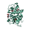 7q8fC  7q8gC 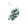 7q8hC 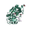 7q8iC  7q8jC  7q8kC  7q8lC  7q8mC 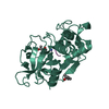 7q8nC  7q8oC  7q8pC  7q9cC  7q9hC  7qffC  7qfhC  7qhjC  7qhkC  1fh0S S: Starting model for refinement C: citing same article ( |
|---|---|
| Similar structure data |
-Links
 PDBj
PDBj


- Assembly
| Deposited unit | 
| ||||||||||||
|---|---|---|---|---|---|---|---|---|---|---|---|---|---|
| 1 | 
| ||||||||||||
| 2 | 
| ||||||||||||
| Unit cell |
| ||||||||||||
| Components on special symmetry positions |
|
-Components
| #1: Protein | / Cathepsin U / Cathepsin V Mass: 24037.936 Da / Num. of mol.: 2 / Mutation: C25S, N179Q Source method: isolated from a genetically manipulated source Source: (gene. exp.) Homo sapiens (human) / Gene: CTSV, CATL2, CTSL2, CTSU, UNQ268/PRO305 / Production host:  Komagataella phaffii GS115 (fungus) / References: UniProt: O60911, cathepsin V Komagataella phaffii GS115 (fungus) / References: UniProt: O60911, cathepsin V#2: Protein/peptide | Mass: 672.817 Da / Num. of mol.: 2 / Source method: obtained synthetically / Source: (synth.) synthetic construct (others) #3: Chemical | 2-Methyl-2,4-pentanediol  Mass: 118.174 Da / Num. of mol.: 3 / Source method: obtained synthetically / Formula: C6H14O2 / Comment: precipitant*YM Mass: 118.174 Da / Num. of mol.: 3 / Source method: obtained synthetically / Formula: C6H14O2 / Comment: precipitant*YM#4: Chemical | Chloride  Mass: 35.453 Da / Num. of mol.: 3 / Source method: obtained synthetically / Formula: Cl Mass: 35.453 Da / Num. of mol.: 3 / Source method: obtained synthetically / Formula: Cl#5: Water | ChemComp-HOH / | Water Mass: 18.015 Da / Num. of mol.: 284 / Source method: isolated from a natural source / Formula: H2O Mass: 18.015 Da / Num. of mol.: 284 / Source method: isolated from a natural source / Formula: H2OHas ligand of interest | N | |
|---|
-Experimental details
-Experiment
| Experiment | Method: X-RAY DIFFRACTION / Number of used crystals: 1 |
|---|
- Sample preparation
| Crystal | Density Matthews: 2.79 Å3/Da / Density % sol: 55.87 % |
|---|---|
| Crystal grow | Temperature: 278 K / Method: vapor diffusion, sitting drop / Details: 77 % MPD, 23 % of 60 mM TRIS, pH 8 |
-Data collection
| Diffraction | Mean temperature: 100 K / Serial crystal experiment: N |
|---|---|
| Diffraction source | Source: SYNCHROTRON / Site: BESSY  / Beamline: 14.1 / Wavelength: 0.9184 Å / Beamline: 14.1 / Wavelength: 0.9184 Å |
| Detector | Type: DECTRIS PILATUS 6M / Detector: PIXEL / Date: Feb 7, 2016 |
| Radiation | Protocol: SINGLE WAVELENGTH / Monochromatic (M) / Laue (L): M / Scattering type: x-ray |
| Radiation wavelength | Wavelength: 0.9184 Å / Relative weight: 1 |
| Reflection | Resolution: 2.13→50 Å / Num. obs: 31532 / % possible obs: 99.3 % / Redundancy: 7.63 % / CC1/2: 0.998 / Net I/σ(I): 14.2 |
| Reflection shell | Resolution: 2.13→2.26 Å / Num. unique obs: 4856 / CC1/2: 0.633 |
- Processing
| Software |
| |||||||||||||||||||||||||
|---|---|---|---|---|---|---|---|---|---|---|---|---|---|---|---|---|---|---|---|---|---|---|---|---|---|---|
| Refinement | Method to determine structure: MOLECULAR REPLACEMENT Starting model: 1FH0 Resolution: 2.13→43.61 Å / Cor.coef. Fo:Fc: 0.8207 / Cor.coef. Fo:Fc free: 0.7723 / Cross valid method: FREE R-VALUE / σ(F): 0 / Phase error: 24.6 / Stereochemistry target values: ML
| |||||||||||||||||||||||||
| Solvent computation | Solvent model: MASK FLAT BULK SOLVENT MODEL / Bsol: 22.94 Å2 / ksol: 0.36 e/Å3 | |||||||||||||||||||||||||
| Displacement parameters | Biso max: 186.73 Å2 / Biso mean: 42.6 Å2 / Biso min: 15.19 Å2
| |||||||||||||||||||||||||
| Refinement step | Cycle: LAST / Resolution: 2.13→43.61 Å
| |||||||||||||||||||||||||
| LS refinement shell | Resolution: 2.13→2.17 Å / Total num. of bins used: 20
|
