- PDB-7q6u: Crystal structure of the bromodomain of ATAD2 with phenol HTS hit... -
+
Open data
ID or keywords:
Loading...
-
Basic information
Entry
Database: PDB / ID: 7q6u
Title
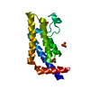
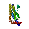
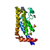
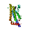
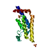
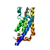
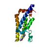
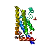
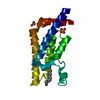
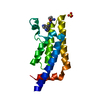
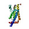
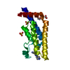
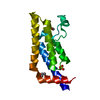
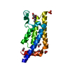
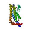
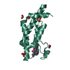
Crystal structure of the bromodomain of ATAD2 with phenol HTS hit (cpd 6)
Components
ATPase family AAA domain-containing protein 2
Keywords
GENE REGULATION / bromodomain / epigenetics
Function / homology
Function and homology information
Hydrolases; Acting on acid anhydrides; In phosphorus-containing anhydrides / TFAP2 (AP-2) family regulates transcription of growth factors and their receptors / histone binding / chromatin binding / positive regulation of DNA-templated transcription / ATP hydrolysis activity / positive regulation of transcription by RNA polymerase II / extracellular exosome / nucleoplasm / ATP binding / nucleus Similarity search - Function
ATPase family AAA domain-containing protein ATAD2-like / AAA ATPase, AAA+ lid domain / AAA+ lid domain / ATPase, AAA-type, conserved site / AAA-protein family signature. / ATPase family associated with various cellular activities (AAA) / ATPase, AAA-type, core / Bromodomain / Bromodomain profile. / bromo domain ...ATPase family AAA domain-containing protein ATAD2-like / AAA ATPase, AAA+ lid domain / AAA+ lid domain / ATPase, AAA-type, conserved site / AAA-protein family signature. / ATPase family associated with various cellular activities (AAA) / ATPase, AAA-type, core / Bromodomain / Bromodomain profile. / bromo domain / Bromodomain / Bromodomain-like superfamily / ATPases associated with a variety of cellular activities / AAA+ ATPase domain / P-loop containing nucleoside triphosphate hydrolase Similarity search - Domain/homology
Mass: 18.015 Da / Num. of mol.: 192 / Source method: isolated from a natural source / Formula: H2O
Has ligand of interest
Y
-
Experimental details
-
Experiment
Experiment
Method: X-RAY DIFFRACTION / Number of used crystals: 1
-
Sample preparation
Crystal
Density Matthews: 4.02 Å3/Da / Density % sol: 69.42 %
Crystal grow
Temperature: 298 K / Method: vapor diffusion / pH: 6.25 Details: ATAD2 (981-1108) at 12 mg/ml in 25 mM Tris pH 9.7, 300 mM NaCl, 0.5 mM TCEP crystallised from 20% PEG 3350, 0.2 M ammonium sulfate, 0.1 M Bis-Tris pH 6.25. Ligands were introduced by ...Details: ATAD2 (981-1108) at 12 mg/ml in 25 mM Tris pH 9.7, 300 mM NaCl, 0.5 mM TCEP crystallised from 20% PEG 3350, 0.2 M ammonium sulfate, 0.1 M Bis-Tris pH 6.25. Ligands were introduced by soaking, cryo-protection with 20 % glycerol.
-
Data collection
Diffraction
Mean temperature: 100 K / Serial crystal experiment: N
In the structure databanks used in Yorodumi, some data are registered as the other names, "COVID-19 virus" and "2019-nCoV". Here are the details of the virus and the list of structure data.
Jan 31, 2019. EMDB accession codes are about to change! (news from PDBe EMDB page)
EMDB accession codes are about to change! (news from PDBe EMDB page)
The allocation of 4 digits for EMDB accession codes will soon come to an end. Whilst these codes will remain in use, new EMDB accession codes will include an additional digit and will expand incrementally as the available range of codes is exhausted. The current 4-digit format prefixed with “EMD-” (i.e. EMD-XXXX) will advance to a 5-digit format (i.e. EMD-XXXXX), and so on. It is currently estimated that the 4-digit codes will be depleted around Spring 2019, at which point the 5-digit format will come into force.
The EM Navigator/Yorodumi systems omit the EMD- prefix.
Related info.:Q: What is EMD? / ID/Accession-code notation in Yorodumi/EM Navigator
Yorodumi is a browser for structure data from EMDB, PDB, SASBDB, etc.
This page is also the successor to EM Navigator detail page, and also detail information page/front-end page for Omokage search.
The word "yorodu" (or yorozu) is an old Japanese word meaning "ten thousand". "mi" (miru) is to see.
Related info.:EMDB / PDB / SASBDB / Comparison of 3 databanks / Yorodumi Search / Aug 31, 2016. New EM Navigator & Yorodumi / Yorodumi Papers / Jmol/JSmol / Function and homology information / Changes in new EM Navigator and Yorodumi
 Movie
Movie Controller

 Yorodumi
 Open data
 Basic information
 Components
 Keywords
 GENE REGULATION /
  Function and homology information
 Authors
 Citation
 Structure visualization
 Downloads & links
 Other downloads
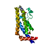


 PDBj
PDBj


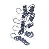
 Assembly


 Mass: 96.063 Da / Num. of mol.: 3 / Source method: obtained synthetically / Formula: SO4
 Mass: 368.383 Da / Num. of mol.: 1 / Source method: obtained synthetically / Formula: C20H20N2O5 / Feature type: SUBJECT OF INVESTIGATION
 Mass: 18.015 Da / Num. of mol.: 192 / Source method: isolated from a natural source / Formula: H2O
 Sample preparation
 Processing