 Movie
Movie Controller

[English] 日本語
 Yorodumi
- PDB-7q3y: Structure of full-length, monomeric, soluble somatic angiotensin ... -
+ Open data

- Basic information
| Entry | Database: PDB / ID: 7q3y | ||||||
|---|---|---|---|---|---|---|---|
| Title | Structure of full-length, monomeric, soluble somatic angiotensin I-converting enzyme showing the N- and C-terminal ellipsoid domains | ||||||
 Components Components | Angiotensin-converting enzyme | ||||||
 Keywords Keywords | HYDROLASE / Zinc metalloprotease Dicarboxypeptidase Glycoprotein | ||||||
| Function / homology |  Function and homology information Function and homology informationmononuclear cell proliferation / cell proliferation in bone marrow / tripeptidyl-peptidase activity / bradykinin receptor binding / regulation of angiotensin metabolic process / exopeptidase activity / substance P catabolic process / response to laminar fluid shear stress / peptidyl-dipeptidase A / regulation of renal output by angiotensin ...mononuclear cell proliferation / cell proliferation in bone marrow / tripeptidyl-peptidase activity / bradykinin receptor binding / regulation of angiotensin metabolic process / exopeptidase activity / substance P catabolic process / response to laminar fluid shear stress / peptidyl-dipeptidase A / regulation of renal output by angiotensin / negative regulation of calcium ion import / positive regulation of peptidyl-cysteine S-nitrosylation / positive regulation of systemic arterial blood pressure / negative regulation of gap junction assembly / metallodipeptidase activity / cellular response to aldosterone / hormone catabolic process / bradykinin catabolic process / angiogenesis involved in coronary vascular morphogenesis / response to thyroid hormone / negative regulation of glucose import / vasoconstriction / neutrophil mediated immunity / hormone metabolic process / regulation of hematopoietic stem cell proliferation / regulation of smooth muscle cell migration / antigen processing and presentation of peptide antigen via MHC class I / mitogen-activated protein kinase binding / embryo development ending in birth or egg hatching / positive regulation of neurogenesis / chloride ion binding / mitogen-activated protein kinase kinase binding / eating behavior / arachidonic acid secretion / post-transcriptional regulation of gene expression / lung alveolus development / peptide catabolic process / heterocyclic compound binding / heart contraction / response to dexamethasone / regulation of heart rate by cardiac conduction / regulation of systemic arterial blood pressure by renin-angiotensin / regulation of vasoconstriction / peptidyl-dipeptidase activity / angiotensin maturation / hematopoietic stem cell differentiation / blood vessel remodeling / Metabolism of Angiotensinogen to Angiotensins / animal organ regeneration / amyloid-beta metabolic process / carboxypeptidase activity / positive regulation of vasoconstriction / sperm midpiece / blood vessel diameter maintenance / response to nutrient levels / basal plasma membrane / kidney development / female pregnancy / angiotensin-activated signaling pathway / cellular response to glucose stimulus / brush border membrane / regulation of synaptic plasticity / metalloendopeptidase activity / regulation of blood pressure / male gonad development / metallopeptidase activity / actin binding / peptidase activity / spermatogenesis / endopeptidase activity / response to lipopolysaccharide / lysosome / response to hypoxia / calmodulin binding / endosome / response to xenobiotic stimulus / positive regulation of apoptotic process / external side of plasma membrane / negative regulation of gene expression / proteolysis / extracellular space / extracellular exosome / zinc ion binding / extracellular region / plasma membraneSimilarity search - Function | ||||||
| Biological species |  Homo sapiens (human) Homo sapiens (human) | ||||||
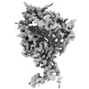
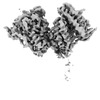
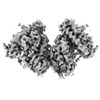
| Method | ELECTRON MICROSCOPY / single particle reconstruction / cryo EM / Resolution: 4.34 Å | ||||||
 Authors Authors | Lubbe, L. / Sewell, B.T. / Sturrock, E.D. | ||||||
| Funding support |  United Kingdom, 1items United Kingdom, 1items
| ||||||
 Citation Citation | Journal: EMBO J / Year: 2022 Title: Cryo-EM reveals mechanisms of angiotensin I-converting enzyme allostery and dimerization. Authors: Lizelle Lubbe / Bryan Trevor Sewell / Jeremy D Woodward / Edward D Sturrock /  Abstract: Hypertension (high blood pressure) is a major risk factor for cardiovascular disease, which is the leading cause of death worldwide. The somatic isoform of angiotensin I-converting enzyme (sACE) ...Hypertension (high blood pressure) is a major risk factor for cardiovascular disease, which is the leading cause of death worldwide. The somatic isoform of angiotensin I-converting enzyme (sACE) plays a critical role in blood pressure regulation, and ACE inhibitors are thus widely used to treat hypertension and cardiovascular disease. Our current understanding of sACE structure, dynamics, function, and inhibition has been limited because truncated, minimally glycosylated forms of sACE are typically used for X-ray crystallography and molecular dynamics simulations. Here, we report the first cryo-EM structures of full-length, glycosylated, soluble sACE (sACE ). Both monomeric and dimeric forms of the highly flexible apo enzyme were reconstructed from a single dataset. The N- and C-terminal domains of monomeric sACE were resolved at 3.7 and 4.1 Å, respectively, while the interacting N-terminal domains responsible for dimer formation were resolved at 3.8 Å. Mechanisms are proposed for intradomain hinging, cooperativity, and homodimerization. Furthermore, the observation that both domains were in the open conformation has implications for the design of sACE modulators. | ||||||
| History |
|
- Structure visualization
| Structure viewer | Molecule: MolmilJmol/JSmol |
|---|
- Downloads & links
-Download
| PDBx/mmCIF format | 7q3y.cif.gz | 430.7 KB | Display | PDBx/mmCIF format |
|---|---|---|---|---|
| PDB format | pdb7q3y.ent.gz | 355 KB | Display | PDB format |
| PDBx/mmJSON format | 7q3y.json.gz | Tree view | PDBx/mmJSON format | |
| Others |  Other downloads Other downloads |
-Validation report
| Arichive directory | https://data.pdbj.org/pub/pdb/validation_reports/q3/7q3yftp://data.pdbj.org/pub/pdb/validation_reports/q3/7q3y | HTTPS FTP |
|---|
-Related structure data
| Related structure data | 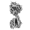 13797MC 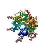 7q49C 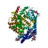 7q4cC 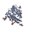 7q4dC 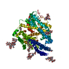 7q4eC M: map data used to model this data C: citing same article ( |
|---|---|
| Similar structure data | |
| EM raw data | EMPIAR-10980 (Title: Cryo-EM structures of monomeric and dimeric human somatic angiotensin I-converting enzyme (apo form) Data size: 3.8 TB Data #1: Unaligned multi-frame cryo-EM micrographs of human somatic angiotensin I-converting enzyme in the apo state [micrographs - multiframe]) |

-Links
 PDBj
PDBj


- Assembly
| Deposited unit | 
|
|---|---|
| 1 |
|
-Components
-Protein , 1 types, 1 molecules A
| #1: Protein | / ACE / Dipeptidyl carboxypeptidase I / Kininase II Mass: 139614.000 Da / Num. of mol.: 1 / Mutation: P576L Source method: isolated from a genetically manipulated source Details: Soluble secreted form of human somatic angiotensin I-converting enzyme terminating at Ser1211 Source: (gene. exp.) Homo sapiens (human) / Gene: ACE, DCP, DCP1 / Plasmid: pcDNA3.1+ / Cell line (production host): CHO-K1 / Production host:   Cricetulus griseus (Chinese hamster) Cricetulus griseus (Chinese hamster)References: UniProt: P12821, Hydrolases; Glycosylases; Glycosidases, i.e. enzymes that hydrolyse O- and S-glycosyl compounds, peptidyl-dipeptidase A |
|---|
-Sugars , 6 types, 13 molecules 
| #2: Polysaccharide | alpha-D-mannopyranose-(1-6)-beta-D-mannopyranose-(1-4)-2-acetamido-2-deoxy-beta-D-glucopyranose-(1- ...alpha-D-mannopyranose-(1-6)-beta-D-mannopyranose-(1-4)-2-acetamido-2-deoxy-beta-D-glucopyranose-(1-4)-2-acetamido-2-deoxy-beta-D-glucopyranose / Mass: 748.682 Da / Num. of mol.: 1 Source method: isolated from a genetically manipulated source | ||||||||
|---|---|---|---|---|---|---|---|---|---|
| #3: Polysaccharide | / Mass: 424.401 Da / Num. of mol.: 2 Source method: isolated from a genetically manipulated source #4: Polysaccharide | beta-D-mannopyranose-(1-4)-2-acetamido-2-deoxy-beta-D-glucopyranose-(1-4)-2-acetamido-2-deoxy-beta- ...beta-D-mannopyranose-(1-4)-2-acetamido-2-deoxy-beta-D-glucopyranose-(1-4)-2-acetamido-2-deoxy-beta-D-glucopyranose / Mass: 586.542 Da / Num. of mol.: 4Source method: isolated from a genetically manipulated source #5: Polysaccharide | alpha-D-mannopyranose-(1-3)-[alpha-D-mannopyranose-(1-6)]beta-D-mannopyranose-(1-4)-2-acetamido-2- ...alpha-D-mannopyranose-(1-3)-[alpha-D-mannopyranose-(1-6)]beta-D-mannopyranose-(1-4)-2-acetamido-2-deoxy-beta-D-glucopyranose-(1-4)-2-acetamido-2-deoxy-beta-D-glucopyranose | / Mass: 910.823 Da / Num. of mol.: 1Source method: isolated from a genetically manipulated source #6: Polysaccharide | alpha-D-mannopyranose-(1-3)-[alpha-D-mannopyranose-(1-6)]beta-D-mannopyranose-(1-4)-2-acetamido-2- ...alpha-D-mannopyranose-(1-3)-[alpha-D-mannopyranose-(1-6)]beta-D-mannopyranose-(1-4)-2-acetamido-2-deoxy-beta-D-glucopyranose-(1-4)-[alpha-L-fucopyranose-(1-6)]2-acetamido-2-deoxy-beta-D-glucopyranose | / Mass: 1056.964 Da / Num. of mol.: 1Source method: isolated from a genetically manipulated source #9: Sugar | ChemComp-NAG / N-Acetylglucosamine Type: D-saccharide, beta linking / Mass: 221.208 Da / Num. of mol.: 4 / Source method: obtained synthetically / Formula: C8H15NO6 / Feature type: SUBJECT OF INVESTIGATION Type: D-saccharide, beta linking / Mass: 221.208 Da / Num. of mol.: 4 / Source method: obtained synthetically / Formula: C8H15NO6 / Feature type: SUBJECT OF INVESTIGATION |
-Non-polymers , 3 types, 3 molecules 


| #7: Chemical | ChemComp-ZN /  Mass: 65.409 Da / Num. of mol.: 1 / Source method: obtained synthetically / Formula: Zn / Feature type: SUBJECT OF INVESTIGATION Mass: 65.409 Da / Num. of mol.: 1 / Source method: obtained synthetically / Formula: Zn / Feature type: SUBJECT OF INVESTIGATION |
|---|---|
| #8: Chemical | ChemComp-CL / Chloride Mass: 35.453 Da / Num. of mol.: 1 / Source method: obtained synthetically / Formula: Cl / Feature type: SUBJECT OF INVESTIGATION Mass: 35.453 Da / Num. of mol.: 1 / Source method: obtained synthetically / Formula: Cl / Feature type: SUBJECT OF INVESTIGATION |
| #10: Water | ChemComp-HOH / WaterMass: 18.015 Da / Num. of mol.: 1 / Source method: isolated from a natural source / Formula: H2O |
-Details
| Has ligand of interest | Y |
|---|
-Experimental details
-Experiment
| Experiment | Method: ELECTRON MICROSCOPY |
|---|---|
| EM experiment | Aggregation state: PARTICLE / 3D reconstruction method: single particle reconstruction |
- Sample preparation
| Component | Name: full-length, soluble, monomeric somatic angiotensin I-converting enzyme Type: COMPLEX / Entity ID: #1 / Source: RECOMBINANT | ||||||||||||||||||||
|---|---|---|---|---|---|---|---|---|---|---|---|---|---|---|---|---|---|---|---|---|---|
| Molecular weight | Value: 0.139 MDa / Experimental value: NO | ||||||||||||||||||||
| Source (natural) | Organism: Homo sapiens (human) | ||||||||||||||||||||
| Source (recombinant) | Organism: Cricetulus griseus (Chinese hamster) / Plasmid: pcDNA3.1+ | ||||||||||||||||||||
| Buffer solution | pH: 7.5 / Details: Solutions were prepared with deionized water | ||||||||||||||||||||
| Buffer component |
| ||||||||||||||||||||
| Specimen | Conc.: 1.5 mg/ml / Embedding applied: NO / Shadowing applied: NO / Staining applied: NO / Vitrification applied: YES Details: The protein was stored at 3.0mg/ml in 50mM HEPES (pH 7.5) and diluted immediately prior to grid preparation | ||||||||||||||||||||
| Specimen support | Grid material: COPPER / Grid mesh size: 200 divisions/in. / Grid type: Quantifoil R2/2 | ||||||||||||||||||||
| Vitrification | Instrument: FEI VITROBOT MARK IV / Cryogen name: ETHANE / Humidity: 100 % / Chamber temperature: 277 K Details: Diluted protein (in buffer containing zinc chloride and sodium chloride) was incubated on ice for 30 minutes after which it was applied to the grid, incubated for 30 seconds, and blotted for ...Details: Diluted protein (in buffer containing zinc chloride and sodium chloride) was incubated on ice for 30 minutes after which it was applied to the grid, incubated for 30 seconds, and blotted for 3 seconds before plunging |
- Electron microscopy imaging
| Experimental equipment |  Model: Titan Krios / Image courtesy: FEI Company |
|---|---|
| Microscopy | Model: FEI TITAN KRIOS |
| Electron gun | Electron source: FIELD EMISSION GUN / Accelerating voltage: 300 kV / Illumination mode: FLOOD BEAM |
| Electron lens | Mode: BRIGHT FIELDBright-field microscopy / Nominal magnification: 81000 X / Nominal defocus max: 3000 nm / Nominal defocus min: 1800 nm / Cs: 2.7 mm |
| Specimen holder | Specimen holder model: FEI TITAN KRIOS AUTOGRID HOLDER |
| Image recording | Average exposure time: 3 sec. / Electron dose: 43 e/Å2 / Film or detector model: GATAN K3 (6k x 4k) / Num. of grids imaged: 1 / Num. of real images: 11628 Details: Images were recorded in super-resolution mode with 40 frames per image |
| Image scans | Width: 5760 / Height: 4092 |
- Processing
| Software |
| |||||||||||||||||||||||||||||||||||||||||||||
|---|---|---|---|---|---|---|---|---|---|---|---|---|---|---|---|---|---|---|---|---|---|---|---|---|---|---|---|---|---|---|---|---|---|---|---|---|---|---|---|---|---|---|---|---|---|---|
| EM software |
| |||||||||||||||||||||||||||||||||||||||||||||
| CTF correction | Details: cryoSPARC patch-based CTF estimation / Type: PHASE FLIPPING AND AMPLITUDE CORRECTION | |||||||||||||||||||||||||||||||||||||||||||||
| Particle selection | Num. of particles selected: 1727045 Details: Topaz-denoising was performed on all curated micrographs, a Topaz model trained on this dataset, and used for picking | |||||||||||||||||||||||||||||||||||||||||||||
| Symmetry | Point symmetry: C1 (asymmetric) | |||||||||||||||||||||||||||||||||||||||||||||
| 3D reconstruction | Resolution: 4.34 Å / Resolution method: FSC 0.143 CUT-OFF / Num. of particles: 121372 / Details: cryoSPARC non-uniform refinement was used / Num. of class averages: 81 / Symmetry type: POINT | |||||||||||||||||||||||||||||||||||||||||||||
| Atomic model building | Protocol: FLEXIBLE FIT / Space: REAL / Target criteria: Correlation coefficient | |||||||||||||||||||||||||||||||||||||||||||||
| Refinement | Cross valid method: NONE Stereochemistry target values: GeoStd + Monomer Library + CDL v1.2 | |||||||||||||||||||||||||||||||||||||||||||||
| Displacement parameters | Biso mean: 131.96 Å2 | |||||||||||||||||||||||||||||||||||||||||||||
| Refine LS restraints |
|
