 Movie
Movie Controller

[English] 日本語
 Yorodumi
- PDB-7pul: Crystal structure of Endoglycosidase E GH20 domain from Enterococ... -
+ Open data

- Basic information
| Entry | Database: PDB / ID: 7pul | ||||||
|---|---|---|---|---|---|---|---|
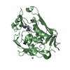
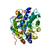
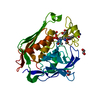
| Title | Crystal structure of Endoglycosidase E GH20 domain from Enterococcus faecalis | ||||||
 Components Components |
| ||||||
 Keywords Keywords |  HYDROLASE / Glycoside hydrolase / GH18 / glycan / antibody / Enterococcus faecalis / EndoE HYDROLASE / Glycoside hydrolase / GH18 / glycan / antibody / Enterococcus faecalis / EndoE | ||||||
| Function / homology |  Function and homology informationbeta-N-acetylhexosaminidase activity / N-acetyl-beta-D-galactosaminidase activity / carbohydrate metabolic process / membrane Function and homology informationbeta-N-acetylhexosaminidase activity / N-acetyl-beta-D-galactosaminidase activity / carbohydrate metabolic process / membraneSimilarity search - Function | ||||||
| Biological species |   Enterococcus faecalis (bacteria) Enterococcus faecalis (bacteria) | ||||||
| Method | X-RAY DIFFRACTION / SYNCHROTRON / MOLECULAR REPLACEMENT / Resolution: 1.4 Å | ||||||
 Authors Authors | Garcia-Alija, M. / Du, J.J. / Trastoy, B. / Sundberg, E.J. / Guerin, M. | ||||||
| Funding support |  United States, 1items United States, 1items
| ||||||
 Citation Citation | Journal: Nat Commun / Year: 2022 Title: Mechanism of cooperative N-glycan processing by the multi-modular endoglycosidase EndoE. Authors: Garcia-Alija, M. / Du, J.J. / Ordonez, I. / Diz-Vallenilla, A. / Moraleda-Montoya, A. / Sultana, N. / Huynh, C.G. / Li, C. / Donahue, T.C. / Wang, L.X. / Trastoy, B. / Sundberg, E.J. / Guerin, M.E. | ||||||
| History |
|
- Structure visualization
| Structure viewer | Molecule: MolmilJmol/JSmol |
|---|
- Downloads & links
-Download
| PDBx/mmCIF format | 7pul.cif.gz | 148.4 KB | Display | PDBx/mmCIF format |
|---|---|---|---|---|
| PDB format | pdb7pul.ent.gz | 114.6 KB | Display | PDB format |
| PDBx/mmJSON format | 7pul.json.gz | Tree view | PDBx/mmJSON format | |
| Others |  Other downloads Other downloads |
-Validation report
| Arichive directory | https://data.pdbj.org/pub/pdb/validation_reports/pu/7pulftp://data.pdbj.org/pub/pdb/validation_reports/pu/7pul | HTTPS FTP |
|---|
-Related structure data
| Related structure data |  7pujC  7pukC 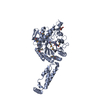 3rpmS S: Starting model for refinement C: citing same article ( |
|---|---|
| Similar structure data |


-Links
 PDBj
PDBj

- Assembly
| Deposited unit | 
| ||||||||
|---|---|---|---|---|---|---|---|---|---|
| 1 |
| ||||||||
| Unit cell |
|
-Components
| #1: Protein | Hexosaminidase Mass: 39906.418 Da / Num. of mol.: 1 Source method: isolated from a genetically manipulated source Source: (gene. exp.) Enterococcus faecalis (bacteria) / Gene: ndoE / Production host: Escherichia coli BL21(DE3) (bacteria) / References: UniProt: Q6U890, beta-N-acetylhexosaminidase | ||||
|---|---|---|---|---|---|
| #2: Protein/peptide | Mass: 345.352 Da / Num. of mol.: 1 Source method: isolated from a genetically manipulated source Source: (gene. exp.) Enterococcus faecalis (bacteria) / Production host: Escherichia coli BL21(DE3) (bacteria) | ||||
| #3: Chemical | ChemComp-CA /   Mass: 40.078 Da / Num. of mol.: 1 / Source method: obtained synthetically / Formula: Ca Mass: 40.078 Da / Num. of mol.: 1 / Source method: obtained synthetically / Formula: Ca | ||||
| #4: Chemical |   Mass: 24.305 Da / Num. of mol.: 3 / Source method: obtained synthetically / Formula: Mg Mass: 24.305 Da / Num. of mol.: 3 / Source method: obtained synthetically / Formula: Mg#5: Water | ChemComp-HOH / | Water Mass: 18.015 Da / Num. of mol.: 309 / Source method: isolated from a natural source / Formula: H2O Mass: 18.015 Da / Num. of mol.: 309 / Source method: isolated from a natural source / Formula: H2OHas ligand of interest | N | |
-Experimental details
-Experiment
| Experiment | Method: X-RAY DIFFRACTION / Number of used crystals: 1 |
|---|
- Sample preparation
| Crystal | Density Matthews: 1.93 Å3/Da / Density % sol: 36.38 % |
|---|---|
| Crystal grow | Temperature: 291 K / Method: vapor diffusion, sitting drop Details: 100 mM bicine/Trizma base pH 8.5, 30 mM magnesium chloride hexahydrate, 30 mM calcium chloride dihydrate, 12.5% w/v PEG 1000, 12.5% PEG w/v 3350, 12.5% v/v MPD |
-Data collection
| Diffraction | Mean temperature: 100 K / Serial crystal experiment: N |
|---|---|
| Diffraction source | Source: SYNCHROTRON / Site: Diamond  / Beamline: I24 / Wavelength: 0.9686 Å / Beamline: I24 / Wavelength: 0.9686 Å |
| Detector | Type: DECTRIS PILATUS3 6M / Detector: PIXEL / Date: Feb 8, 2020 |
| Radiation | Protocol: SINGLE WAVELENGTH / Monochromatic (M) / Laue (L): M / Scattering type: x-ray |
| Radiation wavelength | Wavelength: 0.9686 Å / Relative weight: 1 |
| Reflection | Resolution: 1.4→44.08 Å / Num. obs: 60346 / % possible obs: 99.69 % / Redundancy: 3.2 % / Biso Wilson estimate: 12.65 Å2 / CC1/2: 0.991 / CC star: 0.998 / Rmerge(I) obs: 0.07721 / Rrim(I) all: 0.09335 / Net I/σ(I): 11.92 |
| Reflection shell | Resolution: 1.4→1.45 Å / Redundancy: 3 % / Rmerge(I) obs: 0.2442 / Mean I/σ(I) obs: 2.12 / Num. unique obs: 6021 / CC1/2: 0.894 / CC star: 0.971 / Rrim(I) all: 0.2952 / % possible all: 99.52 |
- Processing
| Software |
| |||||||||||||||||||||||||||||||||||||||||||||||||||||||||||||||||||||||||||||||||||||||||||||||||||||||||||||||||||||||||||||||||||||||||||||||||||||||||||||||||
|---|---|---|---|---|---|---|---|---|---|---|---|---|---|---|---|---|---|---|---|---|---|---|---|---|---|---|---|---|---|---|---|---|---|---|---|---|---|---|---|---|---|---|---|---|---|---|---|---|---|---|---|---|---|---|---|---|---|---|---|---|---|---|---|---|---|---|---|---|---|---|---|---|---|---|---|---|---|---|---|---|---|---|---|---|---|---|---|---|---|---|---|---|---|---|---|---|---|---|---|---|---|---|---|---|---|---|---|---|---|---|---|---|---|---|---|---|---|---|---|---|---|---|---|---|---|---|---|---|---|---|---|---|---|---|---|---|---|---|---|---|---|---|---|---|---|---|---|---|---|---|---|---|---|---|---|---|---|---|---|---|---|---|
| Refinement | Method to determine structure: MOLECULAR REPLACEMENT Starting model: 3RPM Resolution: 1.4→44.077 Å / SU ML: 0.12 / Cross valid method: FREE R-VALUE / σ(F): 1.34 / Phase error: 18.61 / Stereochemistry target values: ML
| |||||||||||||||||||||||||||||||||||||||||||||||||||||||||||||||||||||||||||||||||||||||||||||||||||||||||||||||||||||||||||||||||||||||||||||||||||||||||||||||||
| Solvent computation | Shrinkage radii: 0.9 Å / VDW probe radii: 1.11 Å / Solvent model: FLAT BULK SOLVENT MODEL | |||||||||||||||||||||||||||||||||||||||||||||||||||||||||||||||||||||||||||||||||||||||||||||||||||||||||||||||||||||||||||||||||||||||||||||||||||||||||||||||||
| Refinement step | Cycle: LAST / Resolution: 1.4→44.077 Å
| |||||||||||||||||||||||||||||||||||||||||||||||||||||||||||||||||||||||||||||||||||||||||||||||||||||||||||||||||||||||||||||||||||||||||||||||||||||||||||||||||
| Refine LS restraints |
| |||||||||||||||||||||||||||||||||||||||||||||||||||||||||||||||||||||||||||||||||||||||||||||||||||||||||||||||||||||||||||||||||||||||||||||||||||||||||||||||||
| LS refinement shell |
|
