 Movie
Movie Controller

[English] 日本語
 Yorodumi
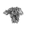
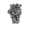
- PDB-7pnq: Human coronavirus OC43 spike glycoprotein ectodomain in complex w... -
+ Open data

- Basic information
| Entry | Database: PDB / ID: 7pnq | |||||||||
|---|---|---|---|---|---|---|---|---|---|---|
| Title | Human coronavirus OC43 spike glycoprotein ectodomain in complex with the 43E6 antibody Fab fragment | |||||||||
 Components Components |
| |||||||||
 Keywords Keywords |  VIRAL PROTEIN / Coronavirus / Glycoprotein / Antibody / Spike VIRAL PROTEIN / Coronavirus / Glycoprotein / Antibody / Spike | |||||||||
| Function / homology |  Function and homology information Function and homology informationendocytosis involved in viral entry into host cell / host cell endoplasmic reticulum-Golgi intermediate compartment membrane / receptor-mediated virion attachment to host cell / fusion of virus membrane with host plasma membrane / fusion of virus membrane with host endosome membrane / viral envelope / host cell plasma membrane / virion membrane / membraneSimilarity search - Function | |||||||||
| Biological species |  Homo sapiens (human) Homo sapiens (human) Human coronavirus OC43 Human coronavirus OC43 | |||||||||
| Method | ELECTRON MICROSCOPY / single particle reconstruction / cryo EM / Resolution: 3.7 Å | |||||||||
 Authors Authors | Hurdiss, D.L. | |||||||||
| Funding support |  Netherlands, Netherlands,  China, 2items China, 2items
| |||||||||
 Citation Citation | Journal: Nat Commun / Year: 2022 Title: Antigenic structure of the human coronavirus OC43 spike reveals exposed and occluded neutralizing epitopes. Authors: Chunyan Wang / Emma L Hesketh / Tatiana M Shamorkina / Wentao Li / Peter J Franken / Dubravka Drabek / Rien van Haperen / Sarah Townend / Frank J M van Kuppeveld / Frank Grosveld / Neil A ...Authors: Chunyan Wang / Emma L Hesketh / Tatiana M Shamorkina / Wentao Li / Peter J Franken / Dubravka Drabek / Rien van Haperen / Sarah Townend / Frank J M van Kuppeveld / Frank Grosveld / Neil A Ranson / Joost Snijder / Raoul J de Groot / Daniel L Hurdiss / Berend-Jan Bosch /  Abstract: Human coronavirus OC43 is a globally circulating common cold virus sustained by recurrent reinfections. How it persists in the population and defies existing herd immunity is unknown. Here we focus ...Human coronavirus OC43 is a globally circulating common cold virus sustained by recurrent reinfections. How it persists in the population and defies existing herd immunity is unknown. Here we focus on viral glycoprotein S, the target for neutralizing antibodies, and provide an in-depth analysis of its antigenic structure. Neutralizing antibodies are directed to the sialoglycan-receptor binding site in S1 domain, but, remarkably, also to S1. The latter block infection yet do not prevent sialoglycan binding. While two distinct neutralizing S1 epitopes are readily accessible in the prefusion S trimer, other sites are occluded such that their accessibility must be subject to conformational changes in S during cell-entry. While non-neutralizing antibodies were broadly reactive against a collection of natural OC43 variants, neutralizing antibodies generally displayed restricted binding breadth. Our data provide a structure-based understanding of protective immunity and adaptive evolution for this endemic coronavirus which emerged in humans long before SARS-CoV-2. | |||||||||
| History |
|
- Structure visualization
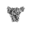
| Structure viewer | Molecule: MolmilJmol/JSmol |
|---|
- Downloads & links
-Download
| PDBx/mmCIF format | 7pnq.cif.gz | 749.1 KB | Display | PDBx/mmCIF format |
|---|---|---|---|---|
| PDB format | pdb7pnq.ent.gz | 596.5 KB | Display | PDB format |
| PDBx/mmJSON format | 7pnq.json.gz | Tree view | PDBx/mmJSON format | |
| Others |  Other downloads Other downloads |
-Validation report
| Arichive directory | https://data.pdbj.org/pub/pdb/validation_reports/pn/7pnqftp://data.pdbj.org/pub/pdb/validation_reports/pn/7pnq | HTTPS FTP |
|---|
-Related structure data
| Related structure data | 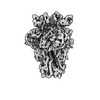 13550MC  7pnmC 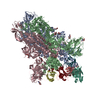 7po5C M: map data used to model this data C: citing same article ( |
|---|---|
| Similar structure data |
-Links
 PDBj
PDBj

- Assembly
| Deposited unit | 
|
|---|---|
| 1 |
|
-Components
| #1: Antibody | Mass: 13511.858 Da / Num. of mol.: 3 Source method: isolated from a genetically manipulated source Source: (gene. exp.) Homo sapiens (human) / Gene: VH / Cell line (production host): HEK293T / Production host: Homo sapiens (human)#2: Antibody | Mass: 11421.707 Da / Num. of mol.: 3 Source method: isolated from a genetically manipulated source Source: (gene. exp.) Homo sapiens (human) / Gene: VL / Cell line (production host): HEK293T / Production host: Homo sapiens (human)#3: Protein | Spike protein / S glycoprotein / E2 / Peplomer proteinMass: 146438.312 Da / Num. of mol.: 3 Source method: isolated from a genetically manipulated source Source: (gene. exp.) Human coronavirus OC43 / Cell line (production host): HEK293T / Production host: Homo sapiens (human) / References: UniProt: Q696P8#4: Sugar | ChemComp-NAG / N-Acetylglucosamine  Type: D-saccharide, beta linking / Mass: 221.208 Da / Num. of mol.: 15 / Source method: obtained synthetically / Formula: C8H15NO6 Type: D-saccharide, beta linking / Mass: 221.208 Da / Num. of mol.: 15 / Source method: obtained synthetically / Formula: C8H15NO6Has ligand of interest | N | |
|---|
-Experimental details
-Experiment
| Experiment | Method: ELECTRON MICROSCOPY |
|---|---|
| EM experiment | Aggregation state: PARTICLE / 3D reconstruction method: single particle reconstruction |
- Sample preparation
| Component | Name: Human coronavirus OC43 spike glycoprotein ectodomain in complex with the 43E6 antibody Fab fragment Type: COMPLEX / Entity ID: #1-#3 / Source: RECOMBINANT | |||||||||||||||
|---|---|---|---|---|---|---|---|---|---|---|---|---|---|---|---|---|
| Molecular weight | Value: 0.583 MDa / Experimental value: NO | |||||||||||||||
| Source (natural) | Organism: Human coronavirus OC43 / Strain: USA/1967 | |||||||||||||||
| Source (recombinant) | Organism: Homo sapiens (human) / Cell: HEK293T | |||||||||||||||
| Buffer solution | pH: 8 | |||||||||||||||
| Buffer component |
| |||||||||||||||
| Specimen | Conc.: 1 mg/ml / Embedding applied: NO / Shadowing applied: NO / Staining applied: NO / Vitrification applied: YES Details: Purified OC43 spike ectodomain and the antibody Fab fragments were incubated together for 5 minutes at a 1:1 molar ratio | |||||||||||||||
| Specimen support | Grid material: COPPER / Grid type: Quantifoil R2/2 | |||||||||||||||
| Vitrification | Instrument: FEI VITROBOT MARK IV / Cryogen name: ETHANE |
- Electron microscopy imaging
| Experimental equipment |  Model: Titan Krios / Image courtesy: FEI Company |
|---|---|
| Microscopy | Model: TFS KRIOS Details: A 30 degree stage tilt was employed during data collection to increase the number of side views visualised due to preferential orientation. |
| Electron gun | Electron source: FIELD EMISSION GUN / Accelerating voltage: 300 kV / Illumination mode: FLOOD BEAM |
| Electron lens | Mode: BRIGHT FIELDBright-field microscopy / Nominal magnification: 75000 X / Nominal defocus max: 2600 nm / Nominal defocus min: 800 nm / Cs: 2.7 mm / C2 aperture diameter: 70 µm |
| Specimen holder | Cryogen: NITROGEN / Specimen holder model: FEI TITAN KRIOS AUTOGRID HOLDER |
| Image recording | Average exposure time: 60 sec. / Electron dose: 52 e/Å2 / Detector mode: COUNTING / Film or detector model: FEI FALCON III (4k x 4k) / Num. of grids imaged: 1 / Num. of real images: 837 |
| Image scans | Width: 4096 / Height: 4096 |
- Processing
| Software |
| ||||||||||||||||||||||||||||||||||||||||
|---|---|---|---|---|---|---|---|---|---|---|---|---|---|---|---|---|---|---|---|---|---|---|---|---|---|---|---|---|---|---|---|---|---|---|---|---|---|---|---|---|---|
| EM software |
| ||||||||||||||||||||||||||||||||||||||||
| CTF correction | Type: PHASE FLIPPING AND AMPLITUDE CORRECTION | ||||||||||||||||||||||||||||||||||||||||
| Particle selection | Num. of particles selected: 349851 | ||||||||||||||||||||||||||||||||||||||||
| Symmetry | Point symmetry: C3 (3 fold cyclic) | ||||||||||||||||||||||||||||||||||||||||
| 3D reconstruction | Resolution: 3.7 Å / Resolution method: FSC 0.143 CUT-OFF / Num. of particles: 53157 / Symmetry type: POINT | ||||||||||||||||||||||||||||||||||||||||
| Atomic model building | PDB-ID: 6NZK | ||||||||||||||||||||||||||||||||||||||||
| Refinement | Cross valid method: NONE Stereochemistry target values: GeoStd + Monomer Library + CDL v1.2 | ||||||||||||||||||||||||||||||||||||||||
| Displacement parameters | Biso mean: 75.82 Å2 | ||||||||||||||||||||||||||||||||||||||||
| Refine LS restraints |
|

