 Movie
Movie Controller

[English] 日本語
 Yorodumi
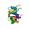
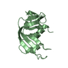
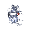
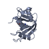
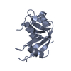
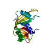
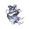
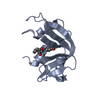
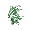
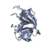
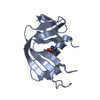
- PDB-7pni: X-ray structure of the adduct formed upon reaction of Pt(II) comp... -
+ Open data

- Basic information
| Entry | Database: PDB / ID: 7pni | ||||||
|---|---|---|---|---|---|---|---|
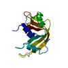
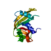
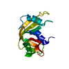
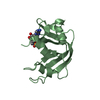
| Title | X-ray structure of the adduct formed upon reaction of Pt(II) complex 2c with ribonuclease A | ||||||
 Components Components | Ribonuclease pancreatic Pancreatic ribonuclease family Pancreatic ribonuclease family | ||||||
 Keywords Keywords | RNA BINDING PROTEIN / metallodrug / platinum / protein interaction / Pt-protein adduct | ||||||
| Function / homology |  Function and homology informationpancreatic ribonuclease / ribonuclease A activity / RNA nuclease activity / nucleic acid binding / lyase activity / defense response to Gram-positive bacterium / extracellular region Function and homology informationpancreatic ribonuclease / ribonuclease A activity / RNA nuclease activity / nucleic acid binding / lyase activity / defense response to Gram-positive bacterium / extracellular regionSimilarity search - Function | ||||||
| Biological species |  Bos taurus (cattle) Bos taurus (cattle) | ||||||
| Method | X-RAY DIFFRACTION / SYNCHROTRON / MOLECULAR REPLACEMENT / Resolution: 2.13 Å | ||||||
 Authors Authors | Ferraro, G. / Merlino, A. | ||||||
| Funding support | 1items
| ||||||
 Citation Citation | Journal: Int J Mol Sci / Year: 2021 Title: Reactions with Proteins of Three Novel Anticancer Platinum(II) Complexes Bearing N-Heterocyclic Ligands. Authors: Sacco, F. / Tarchi, M. / Ferraro, G. / Merlino, A. / Facchetti, G. / Rimoldi, I. / Messori, L. / Massai, L. | ||||||
| History |
|
- Structure visualization
| Structure viewer | Molecule: MolmilJmol/JSmol |
|---|
- Downloads & links
-Download
| PDBx/mmCIF format | 7pni.cif.gz | 68.6 KB | Display | PDBx/mmCIF format |
|---|---|---|---|---|
| PDB format | pdb7pni.ent.gz | Display | PDB format | |
| PDBx/mmJSON format | 7pni.json.gz | Tree view | PDBx/mmJSON format | |
| Others |  Other downloads Other downloads |
-Validation report
| Arichive directory | https://data.pdbj.org/pub/pdb/validation_reports/pn/7pniftp://data.pdbj.org/pub/pdb/validation_reports/pn/7pni | HTTPS FTP |
|---|
-Related structure data
| Related structure data | 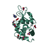 7pnhC  1jvtS S: Starting model for refinement C: citing same article ( |
|---|---|
| Similar structure data |


-Links
 PDBj
PDBj


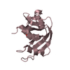
- Assembly
| Deposited unit | 
| ||||||||
|---|---|---|---|---|---|---|---|---|---|
| 1 | 
| ||||||||
| 2 | 
| ||||||||
| Unit cell |
|
-Components
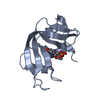
| #1: Protein | Pancreatic ribonuclease family / RNase 1 / RNase A Mass: 13708.326 Da / Num. of mol.: 2 / Source method: isolated from a natural source / Source: (natural) Bos taurus (cattle) / References: UniProt: P61823, pancreatic ribonuclease#2: Chemical | ChemComp-PT /   Mass: 195.078 Da / Num. of mol.: 5 / Source method: obtained synthetically / Formula: Pt / Feature type: SUBJECT OF INVESTIGATION Mass: 195.078 Da / Num. of mol.: 5 / Source method: obtained synthetically / Formula: Pt / Feature type: SUBJECT OF INVESTIGATION#3: Water | ChemComp-HOH / | Water Mass: 18.015 Da / Num. of mol.: 139 / Source method: isolated from a natural source / Formula: H2O Mass: 18.015 Da / Num. of mol.: 139 / Source method: isolated from a natural source / Formula: H2OHas ligand of interest | Y | |
|---|
-Experimental details
-Experiment
| Experiment | Method: X-RAY DIFFRACTION / Number of used crystals: 1 |
|---|
- Sample preparation
| Crystal | Density Matthews: 2.14 Å3/Da / Density % sol: 42.54 % |
|---|---|
| Crystal grow | Temperature: 293 K / Method: vapor diffusion, hanging drop / pH: 5.1 / Details: 22% PEG4K 10 mM sodium citrate pH 5.1 |
-Data collection
| Diffraction | Mean temperature: 100 K / Serial crystal experiment: N |
|---|---|
| Diffraction source | Source: SYNCHROTRON / Site: ELETTRA  / Beamline: 11.2C / Wavelength: 0.96 Å / Beamline: 11.2C / Wavelength: 0.96 Å |
| Detector | Type: DECTRIS PILATUS 6M / Detector: PIXEL / Date: Mar 4, 2021 |
| Radiation | Protocol: SINGLE WAVELENGTH / Monochromatic (M) / Laue (L): M / Scattering type: x-ray |
| Radiation wavelength | Wavelength: 0.96 Å / Relative weight: 1 |
| Reflection | Resolution: 2.13→28.04 Å / Num. obs: 13153 / % possible obs: 99 % / Redundancy: 6.2 % / CC1/2: 0.998 / Rmerge(I) obs: 0.078 / Rpim(I) all: 0.034 / Rrim(I) all: 0.086 / Net I/σ(I): 14.5 |
| Reflection shell | Resolution: 2.13→2.17 Å / Redundancy: 4.9 % / Rmerge(I) obs: 0.643 / Mean I/σ(I) obs: 2.3 / Num. unique obs: 649 / CC1/2: 0.898 / Rpim(I) all: 0.318 / Rrim(I) all: 0.721 / % possible all: 97.3 |
- Processing
| Software |
| |||||||||||||||||||||||||||||||||||||||||||||||||||||||||||||||||||||||||||||||||||||||||||||||||||||||||||||||||||||||||||||||||||||||||||||||||||||||||||||||||||||||||||||||||||||||||||||||||||||||||||||||||||||||||||||||||||||||
|---|---|---|---|---|---|---|---|---|---|---|---|---|---|---|---|---|---|---|---|---|---|---|---|---|---|---|---|---|---|---|---|---|---|---|---|---|---|---|---|---|---|---|---|---|---|---|---|---|---|---|---|---|---|---|---|---|---|---|---|---|---|---|---|---|---|---|---|---|---|---|---|---|---|---|---|---|---|---|---|---|---|---|---|---|---|---|---|---|---|---|---|---|---|---|---|---|---|---|---|---|---|---|---|---|---|---|---|---|---|---|---|---|---|---|---|---|---|---|---|---|---|---|---|---|---|---|---|---|---|---|---|---|---|---|---|---|---|---|---|---|---|---|---|---|---|---|---|---|---|---|---|---|---|---|---|---|---|---|---|---|---|---|---|---|---|---|---|---|---|---|---|---|---|---|---|---|---|---|---|---|---|---|---|---|---|---|---|---|---|---|---|---|---|---|---|---|---|---|---|---|---|---|---|---|---|---|---|---|---|---|---|---|---|---|---|---|---|---|---|---|---|---|---|---|---|---|---|---|---|---|---|---|
| Refinement | Method to determine structure: MOLECULAR REPLACEMENT Starting model: 1jvt Resolution: 2.13→28.04 Å / Cor.coef. Fo:Fc: 0.95 / Cor.coef. Fo:Fc free: 0.903 / SU B: 7.018 / SU ML: 0.18 / Cross valid method: FREE R-VALUE / ESU R: 0.296 / ESU R Free: 0.245 Details: Hydrogens have been added in their riding positions
| |||||||||||||||||||||||||||||||||||||||||||||||||||||||||||||||||||||||||||||||||||||||||||||||||||||||||||||||||||||||||||||||||||||||||||||||||||||||||||||||||||||||||||||||||||||||||||||||||||||||||||||||||||||||||||||||||||||||
| Solvent computation | Ion probe radii: 0.8 Å / Shrinkage radii: 0.8 Å / VDW probe radii: 1.2 Å / Solvent model: MASK BULK SOLVENT | |||||||||||||||||||||||||||||||||||||||||||||||||||||||||||||||||||||||||||||||||||||||||||||||||||||||||||||||||||||||||||||||||||||||||||||||||||||||||||||||||||||||||||||||||||||||||||||||||||||||||||||||||||||||||||||||||||||||
| Displacement parameters | Biso mean: 39.806 Å2
| |||||||||||||||||||||||||||||||||||||||||||||||||||||||||||||||||||||||||||||||||||||||||||||||||||||||||||||||||||||||||||||||||||||||||||||||||||||||||||||||||||||||||||||||||||||||||||||||||||||||||||||||||||||||||||||||||||||||
| Refinement step | Cycle: LAST / Resolution: 2.13→28.04 Å
| |||||||||||||||||||||||||||||||||||||||||||||||||||||||||||||||||||||||||||||||||||||||||||||||||||||||||||||||||||||||||||||||||||||||||||||||||||||||||||||||||||||||||||||||||||||||||||||||||||||||||||||||||||||||||||||||||||||||
| Refine LS restraints |
| |||||||||||||||||||||||||||||||||||||||||||||||||||||||||||||||||||||||||||||||||||||||||||||||||||||||||||||||||||||||||||||||||||||||||||||||||||||||||||||||||||||||||||||||||||||||||||||||||||||||||||||||||||||||||||||||||||||||
| LS refinement shell | Refine-ID: X-RAY DIFFRACTION / Total num. of bins used: 20
|
