 Movie
Movie Controller

[English] 日本語
 Yorodumi
- PDB-7pc5: The third PDZ domain of PDZD7 complexed with the PDZ-binding moti... -
+ Open data

- Basic information
| Entry | Database: PDB / ID: 7pc5 | ||||||
|---|---|---|---|---|---|---|---|
| Title | The third PDZ domain of PDZD7 complexed with the PDZ-binding motif of EXOC4 | ||||||
 Components Components |
| ||||||
 Keywords Keywords | PEPTIDE BINDING PROTEIN / PDZ /  complex / crystallization chaperone complex / crystallization chaperone | ||||||
| Function / homology |  Function and homology information Function and homology informationvesicle tethering involved in exocytosis / paraxial mesoderm formation / USH2 complex / stereocilia ankle link / inner ear receptor cell differentiation / stereocilia ankle link complex / exocyst / positive regulation of low-density lipoprotein particle receptor binding / positive regulation of receptor-mediated endocytosis involved in cholesterol transport / AnxA2-p11 complex ...vesicle tethering involved in exocytosis / paraxial mesoderm formation / USH2 complex / stereocilia ankle link / inner ear receptor cell differentiation / stereocilia ankle link complex / exocyst / positive regulation of low-density lipoprotein particle receptor binding / positive regulation of receptor-mediated endocytosis involved in cholesterol transport / AnxA2-p11 complex / membrane raft assembly / protein localization to ciliary membrane / positive regulation of vacuole organization / positive regulation of low-density lipoprotein particle clearance / phospholipase A2 inhibitor activity / positive regulation of vesicle fusion / negative regulation of low-density lipoprotein particle receptor catabolic process / positive regulation of plasma membrane repair / positive regulation of plasminogen activation / stereocilium tip / PCSK9-AnxA2 complex / growth cone membrane / : / VxPx cargo-targeting to cilium / myelin sheath abaxonal region / myelin sheath adaxonal region / auditory receptor cell development / detection of mechanical stimulus involved in sensory perception of sound / cadherin binding involved in cell-cell adhesion / Schmidt-Lanterman incisure / Golgi to plasma membrane transport / vesicle budding from membrane / stereocilium / cornified envelope / vesicle docking involved in exocytosis / plasma membrane protein complex / calcium-dependent phospholipid binding / negative regulation of receptor internalization / Flemming body / collagen fibril organization / auditory receptor cell stereocilium organization / S100 protein binding / Dissolution of Fibrin Clot / virion binding / osteoclast development / positive regulation of low-density lipoprotein receptor activity / epithelial cell apoptotic process / phosphatidylserine binding / positive regulation of receptor recycling / Insulin processing / exocytosis / positive regulation of exocytosis / microvillus / cilium assembly / basement membrane / regulation of neurogenesis / Smooth Muscle Contraction / regulation of macroautophagy / fibrinolysis / phosphatidylinositol-4,5-bisphosphate binding / Gene and protein expression by JAK-STAT signaling after Interleukin-12 stimulation / cytoskeletal protein binding / lipid droplet / cell-matrix adhesion / response to activity / Translocation of SLC2A4 (GLUT4) to the plasma membrane / establishment of localization in cell / PDZ domain binding / sensory perception of sound / adherens junction / lung development / sarcolemma / mRNA transcription by RNA polymerase II / serine-type endopeptidase inhibitor activity / establishment of protein localization / calcium channel activity / cilium / small GTPase binding / nuclear matrix / RNA polymerase II transcription regulator complex / calcium-dependent protein binding / azurophil granule lumen / melanosome / protein transport / late endosome membrane / midbody / chemical synaptic transmission / basolateral plasma membrane / angiogenesis / collagen-containing extracellular matrix / protease binding / vesicle / early endosome / endosome / lysosomal membrane / synapse / calcium ion binding / Neutrophil degranulation / cell surface / positive regulation of transcription by RNA polymerase IISimilarity search - Function | ||||||
| Biological species |  Homo sapiens (human) Homo sapiens (human) | ||||||
| Method | X-RAY DIFFRACTION / SYNCHROTRON / MOLECULAR REPLACEMENT / Resolution: 1.7 Å | ||||||
 Authors Authors | Cousido-Siah, A. / Trave, G. / Gogl, G. | ||||||
| Funding support | 1items
| ||||||
 Citation Citation | Journal: Acta Crystallogr D Struct Biol / Year: 2022 Title: A scalable strategy to solve structures of PDZ domains and their complexes. Authors: Cousido-Siah, A. / Carneiro, L. / Kostmann, C. / Ecsedi, P. / Nyitray, L. / Trave, G. / Gogl, G. | ||||||
| History |
|
- Structure visualization
| Structure viewer | Molecule: MolmilJmol/JSmol |
|---|
- Downloads & links
-Download
| PDBx/mmCIF format | 7pc5.cif.gz | 196.9 KB | Display | PDBx/mmCIF format |
|---|---|---|---|---|
| PDB format | pdb7pc5.ent.gz | 153.6 KB | Display | PDB format |
| PDBx/mmJSON format | 7pc5.json.gz | Tree view | PDBx/mmJSON format | |
| Others |  Other downloads Other downloads |
-Validation report
| Arichive directory | https://data.pdbj.org/pub/pdb/validation_reports/pc/7pc5ftp://data.pdbj.org/pub/pdb/validation_reports/pc/7pc5 | HTTPS FTP |
|---|
-Related structure data
| Related structure data |  7pc3C 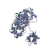 7pc4C 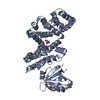 7pc7C 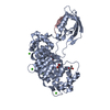 7pc8C 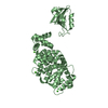 7pc9C 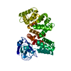 7pcbC  7qqlC 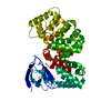 7qqmC  7qqnC  3lnyS  5n7dS S: Starting model for refinement C: citing same article ( |
|---|---|
| Similar structure data |
-Links
 PDBj
PDBj


- Assembly
| Deposited unit | 
| ||||||||
|---|---|---|---|---|---|---|---|---|---|
| 1 |
| ||||||||
| Unit cell |
|
-Components
| #1: Protein | Mass: 47297.020 Da / Num. of mol.: 1 Source method: isolated from a genetically manipulated source Source: (gene. exp.) Homo sapiens (human) / Gene: PDZD7, PDZK7, ANXA2, ANX2, ANX2L4, CAL1H, LPC2D / Production host:  Escherichia coli (E. coli) / References: UniProt: Q9H5P4, UniProt: P07355 Escherichia coli (E. coli) / References: UniProt: Q9H5P4, UniProt: P07355 | ||||||
|---|---|---|---|---|---|---|---|
| #2: Protein/peptide | Exocyst / Exocyst complex component Sec8 Mass: 1107.320 Da / Num. of mol.: 1 / Source method: obtained synthetically Details: N-terminal biotin-ado-ado (amino-dodecanoic acid) label Source: (synth.) Homo sapiens (human) / References: UniProt: Q96A65 | ||||||
| #3: Chemical | ChemComp-CA /   Mass: 40.078 Da / Num. of mol.: 5 / Source method: obtained synthetically / Formula: Ca Mass: 40.078 Da / Num. of mol.: 5 / Source method: obtained synthetically / Formula: Ca#4: Chemical | Glycerol  Mass: 92.094 Da / Num. of mol.: 2 / Source method: obtained synthetically / Formula: C3H8O3 Mass: 92.094 Da / Num. of mol.: 2 / Source method: obtained synthetically / Formula: C3H8O3#5: Water | ChemComp-HOH / | Water Mass: 18.015 Da / Num. of mol.: 504 / Source method: isolated from a natural source / Formula: H2O Mass: 18.015 Da / Num. of mol.: 504 / Source method: isolated from a natural source / Formula: H2OHas ligand of interest | N | |
-Experimental details
-Experiment
| Experiment | Method: X-RAY DIFFRACTION / Number of used crystals: 1 |
|---|
- Sample preparation
| Crystal | Density Matthews: 3.18 Å3/Da / Density % sol: 61.28 % |
|---|---|
| Crystal grow | Temperature: 293 K / Method: vapor diffusion, sitting drop Details: 0.2M ammonium sulfate, 15% PEG 4000, 0.1M TRIS pH8.5 |
-Data collection
| Diffraction | Mean temperature: 100 K / Serial crystal experiment: N |
|---|---|
| Diffraction source | Source: SYNCHROTRON / Site: SLS  / Beamline: X06DA / Wavelength: 1 Å / Beamline: X06DA / Wavelength: 1 Å |
| Detector | Type: DECTRIS PILATUS 2M / Detector: PIXEL / Date: Jun 14, 2021 |
| Radiation | Protocol: SINGLE WAVELENGTH / Monochromatic (M) / Laue (L): M / Scattering type: x-ray |
| Radiation wavelength | Wavelength: 1 Å / Relative weight: 1 |
| Reflection | Resolution: 1.7→42.748 Å / Num. obs: 64942 / % possible obs: 97.6 % / Redundancy: 13.64 % / CC1/2: 0.999 / Rrim(I) all: 0.1 / Net I/σ(I): 16.64 |
| Reflection shell | Resolution: 1.7→1.74 Å / Mean I/σ(I) obs: 1.83 / Num. unique obs: 4670 / CC1/2: 0.715 / Rrim(I) all: 1.561 |
- Processing
| Software |
| ||||||||||||||||||||||||||||||||||||||||||||||||||||||||||||||||||||||||||||||||||||||||||||||||||||||||||||||||||||||||||||||||||||||||||||||||
|---|---|---|---|---|---|---|---|---|---|---|---|---|---|---|---|---|---|---|---|---|---|---|---|---|---|---|---|---|---|---|---|---|---|---|---|---|---|---|---|---|---|---|---|---|---|---|---|---|---|---|---|---|---|---|---|---|---|---|---|---|---|---|---|---|---|---|---|---|---|---|---|---|---|---|---|---|---|---|---|---|---|---|---|---|---|---|---|---|---|---|---|---|---|---|---|---|---|---|---|---|---|---|---|---|---|---|---|---|---|---|---|---|---|---|---|---|---|---|---|---|---|---|---|---|---|---|---|---|---|---|---|---|---|---|---|---|---|---|---|---|---|---|---|---|---|
| Refinement | Method to determine structure: MOLECULAR REPLACEMENT Starting model: 5N7D, 3LNY Resolution: 1.7→42.748 Å / SU ML: 0.17 / Cross valid method: THROUGHOUT / σ(F): 1.36 / Phase error: 18.78 / Stereochemistry target values: ML
| ||||||||||||||||||||||||||||||||||||||||||||||||||||||||||||||||||||||||||||||||||||||||||||||||||||||||||||||||||||||||||||||||||||||||||||||||
| Solvent computation | Shrinkage radii: 1 Å / VDW probe radii: 1.2 Å / Solvent model: FLAT BULK SOLVENT MODEL | ||||||||||||||||||||||||||||||||||||||||||||||||||||||||||||||||||||||||||||||||||||||||||||||||||||||||||||||||||||||||||||||||||||||||||||||||
| Displacement parameters | Biso max: 130.68 Å2 / Biso mean: 40.7018 Å2 / Biso min: 13.93 Å2 | ||||||||||||||||||||||||||||||||||||||||||||||||||||||||||||||||||||||||||||||||||||||||||||||||||||||||||||||||||||||||||||||||||||||||||||||||
| Refinement step | Cycle: final / Resolution: 1.7→42.748 Å
| ||||||||||||||||||||||||||||||||||||||||||||||||||||||||||||||||||||||||||||||||||||||||||||||||||||||||||||||||||||||||||||||||||||||||||||||||
| LS refinement shell | Refine-ID: X-RAY DIFFRACTION / Rfactor Rfree error: 0
| ||||||||||||||||||||||||||||||||||||||||||||||||||||||||||||||||||||||||||||||||||||||||||||||||||||||||||||||||||||||||||||||||||||||||||||||||
| Refinement TLS params. | Method: refined / Refine-ID: X-RAY DIFFRACTION
| ||||||||||||||||||||||||||||||||||||||||||||||||||||||||||||||||||||||||||||||||||||||||||||||||||||||||||||||||||||||||||||||||||||||||||||||||
| Refinement TLS group |
|
