 Movie
Movie Controller

[English] 日本語
 Yorodumi
- PDB-7p84: Crystal structure of L147A/I351A variant of S-adenosylmethionine ... -
+ Open data

- Basic information
| Entry | Database: PDB / ID: 7p84 | ||||||
|---|---|---|---|---|---|---|---|
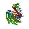
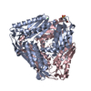
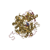
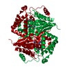
| Title | Crystal structure of L147A/I351A variant of S-adenosylmethionine synthetase from Methanocaldococcus jannaschii in complex with ONB-SAM (2-nitro benzyme S-adenosyl-methionine) | ||||||
 Components Components | S-adenosylmethionine synthase | ||||||
 Keywords Keywords |  TRANSFERASE TRANSFERASE | ||||||
| Function / homology |  Function and homology informationmethionine adenosyltransferase / methionine adenosyltransferase activity / S-adenosylmethionine biosynthetic process / one-carbon metabolic process / magnesium ion binding / ATP binding Function and homology informationmethionine adenosyltransferase / methionine adenosyltransferase activity / S-adenosylmethionine biosynthetic process / one-carbon metabolic process / magnesium ion binding / ATP bindingSimilarity search - Function | ||||||
| Biological species |   Methanocaldococcus jannaschii (archaea) Methanocaldococcus jannaschii (archaea) | ||||||
| Method | X-RAY DIFFRACTION / SYNCHROTRON / MOLECULAR REPLACEMENT / Resolution: 2.054 Å | ||||||
 Authors Authors | Herrmann, E. / Peters, A. / Cornelissen, N.V. / Rentmeister, A. / Kuemmel, D. | ||||||
 Citation Citation | Journal: Chembiochem / Year: 2022 Title: Visible-Light Removable Photocaging Groups Accepted by MjMAT Variant: Structural Basis and Compatibility with DNA and RNA Methyltransferases. Authors: Peters, A. / Herrmann, E. / Cornelissen, N.V. / Klocker, N. / Kummel, D. / Rentmeister, A. | ||||||
| History |
|
- Structure visualization
| Structure viewer | Molecule: MolmilJmol/JSmol |
|---|
- Downloads & links
-Download
| PDBx/mmCIF format | 7p84.cif.gz | 319.8 KB | Display | PDBx/mmCIF format |
|---|---|---|---|---|
| PDB format | pdb7p84.ent.gz | 259.7 KB | Display | PDB format |
| PDBx/mmJSON format | 7p84.json.gz | Tree view | PDBx/mmJSON format | |
| Others |  Other downloads Other downloads |
-Validation report
| Arichive directory | https://data.pdbj.org/pub/pdb/validation_reports/p8/7p84ftp://data.pdbj.org/pub/pdb/validation_reports/p8/7p84 | HTTPS FTP |
|---|
-Related structure data
| Related structure data |  7p82SC  7p83C  7p8mC S: Starting model for refinement C: citing same article ( |
|---|---|
| Similar structure data |


-Links
 PDBj
PDBj

- Assembly
| Deposited unit | 
| ||||||||
|---|---|---|---|---|---|---|---|---|---|
| 1 |
| ||||||||
| Unit cell |
|
-Components
| #1: Protein | Mass: 47401.336 Da / Num. of mol.: 2 / Mutation: L147A/I351A Source method: isolated from a genetically manipulated source Source: (gene. exp.) Methanocaldococcus jannaschii (strain ATCC 43067 / DSM 2661 / JAL-1 / JCM 10045 / NBRC 100440) (archaea)Gene: mat, MJ1208 / Production host:  Escherichia coli BL21(DE3) (bacteria) / References: UniProt: Q58605, methionine adenosyltransferase Escherichia coli BL21(DE3) (bacteria) / References: UniProt: Q58605, methionine adenosyltransferase#2: Chemical | Polyphosphate  Mass: 257.955 Da / Num. of mol.: 2 / Source method: obtained synthetically / Formula: H5O10P3 Mass: 257.955 Da / Num. of mol.: 2 / Source method: obtained synthetically / Formula: H5O10P3#3: Chemical |   Mass: 24.305 Da / Num. of mol.: 3 / Source method: obtained synthetically / Formula: Mg Mass: 24.305 Da / Num. of mol.: 3 / Source method: obtained synthetically / Formula: Mg#4: Chemical | ChemComp-EU9 / [( |   Mass: 520.539 Da / Num. of mol.: 1 / Source method: obtained synthetically / Formula: C21H26N7O7S / Feature type: SUBJECT OF INVESTIGATION Mass: 520.539 Da / Num. of mol.: 1 / Source method: obtained synthetically / Formula: C21H26N7O7S / Feature type: SUBJECT OF INVESTIGATION#5: Water | ChemComp-HOH / | Water Mass: 18.015 Da / Num. of mol.: 387 / Source method: isolated from a natural source / Formula: H2O Mass: 18.015 Da / Num. of mol.: 387 / Source method: isolated from a natural source / Formula: H2OHas ligand of interest | Y | |
|---|
-Experimental details
-Experiment
| Experiment | Method: X-RAY DIFFRACTION / Number of used crystals: 1 |
|---|
- Sample preparation
| Crystal | Density Matthews: 2.59 Å3/Da / Density % sol: 52.46 % |
|---|---|
| Crystal grow | Temperature: 293.15 K / Method: vapor diffusion, sitting drop / pH: 4.6 / Details: 0.1 M sodium acetate pH 4.6, 40% (v/v) PEG200 |
-Data collection
| Diffraction | Mean temperature: 100 K / Serial crystal experiment: N |
|---|---|
| Diffraction source | Source: SYNCHROTRON / Site: BESSY  / Beamline: 14.1 / Wavelength: 0.9184 Å / Beamline: 14.1 / Wavelength: 0.9184 Å |
| Detector | Type: DECTRIS PILATUS3 S 6M / Detector: PIXEL / Date: Feb 3, 2021 |
| Radiation | Protocol: SINGLE WAVELENGTH / Monochromatic (M) / Laue (L): M / Scattering type: x-ray |
| Radiation wavelength | Wavelength: 0.9184 Å / Relative weight: 1 |
| Reflection | Resolution: 2.05→48.04 Å / Num. obs: 112614 / % possible obs: 99.3 % / Redundancy: 5.59 % / CC1/2: 0.996 / Rrim(I) all: 0.155 / Net I/σ(I): 8.1 |
| Reflection shell | Resolution: 2.05→2.18 Å / Mean I/σ(I) obs: 1.58 / Num. unique obs: 17696 / CC1/2: 0.919 / Rrim(I) all: 0.968 |
- Processing
| Software |
| ||||||||||||||||||||||||||||||||||||||||||||||||||||||||||||||||||||||||||||||||||||||||||||||||
|---|---|---|---|---|---|---|---|---|---|---|---|---|---|---|---|---|---|---|---|---|---|---|---|---|---|---|---|---|---|---|---|---|---|---|---|---|---|---|---|---|---|---|---|---|---|---|---|---|---|---|---|---|---|---|---|---|---|---|---|---|---|---|---|---|---|---|---|---|---|---|---|---|---|---|---|---|---|---|---|---|---|---|---|---|---|---|---|---|---|---|---|---|---|---|---|---|---|
| Refinement | Method to determine structure: MOLECULAR REPLACEMENT Starting model: 7P82 Resolution: 2.054→48.04 Å / SU ML: 0.25 / Cross valid method: THROUGHOUT / σ(F): 1.34 / Phase error: 23 / Stereochemistry target values: ML
| ||||||||||||||||||||||||||||||||||||||||||||||||||||||||||||||||||||||||||||||||||||||||||||||||
| Solvent computation | Shrinkage radii: 0.9 Å / VDW probe radii: 1.11 Å / Solvent model: FLAT BULK SOLVENT MODEL | ||||||||||||||||||||||||||||||||||||||||||||||||||||||||||||||||||||||||||||||||||||||||||||||||
| Displacement parameters | Biso max: 151.43 Å2 / Biso mean: 38.66 Å2 / Biso min: 17.74 Å2 | ||||||||||||||||||||||||||||||||||||||||||||||||||||||||||||||||||||||||||||||||||||||||||||||||
| Refinement step | Cycle: final / Resolution: 2.054→48.04 Å
| ||||||||||||||||||||||||||||||||||||||||||||||||||||||||||||||||||||||||||||||||||||||||||||||||
| Refine LS restraints |
| ||||||||||||||||||||||||||||||||||||||||||||||||||||||||||||||||||||||||||||||||||||||||||||||||
| LS refinement shell | Refine-ID: X-RAY DIFFRACTION / Rfactor Rfree error: 0
|
