 Movie
Movie Controller


+ Open data

- Basic information
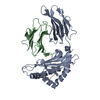
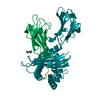
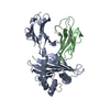
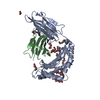
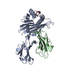
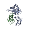
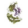
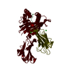
| Entry | Database: PDB / ID: 7p3d | ||||||
|---|---|---|---|---|---|---|---|
| Title | MHC I A02 Allele presenting YLQPRTFLL | ||||||
 Components Components |
| ||||||
 Keywords Keywords |  IMMUNE SYSTEM / Viral Epitope / Wuhan virus / S protein epitope / MHC presentation IMMUNE SYSTEM / Viral Epitope / Wuhan virus / S protein epitope / MHC presentation | ||||||
| Function / homology |  Function and homology information Function and homology informationantigen processing and presentation of endogenous peptide antigen via MHC class I via ER pathway, TAP-independent / antigen processing and presentation of endogenous peptide antigen via MHC class Ib / positive regulation of ferrous iron binding / positive regulation of transferrin receptor binding / positive regulation of receptor binding / early endosome lumen / Nef mediated downregulation of MHC class I complex cell surface expression / DAP12 interactions / negative regulation of receptor binding / Endosomal/Vacuolar pathway ...antigen processing and presentation of endogenous peptide antigen via MHC class I via ER pathway, TAP-independent / antigen processing and presentation of endogenous peptide antigen via MHC class Ib / positive regulation of ferrous iron binding / positive regulation of transferrin receptor binding / positive regulation of receptor binding / early endosome lumen / Nef mediated downregulation of MHC class I complex cell surface expression / DAP12 interactions / negative regulation of receptor binding / Endosomal/Vacuolar pathway / Antigen Presentation: Folding, assembly and peptide loading of class I MHC / cellular response to iron(III) ion / antigen processing and presentation of exogenous protein antigen via MHC class Ib, TAP-dependent / negative regulation of forebrain neuron differentiation / ER to Golgi transport vesicle membrane / regulation of erythrocyte differentiation / peptide antigen assembly with MHC class I protein complex / response to molecule of bacterial origin / regulation of iron ion transport / MHC class I peptide loading complex / HFE-transferrin receptor complex / T cell mediated cytotoxicity / cellular response to iron ion / antigen processing and presentation of endogenous peptide antigen via MHC class I / positive regulation of T cell cytokine production / MHC class I protein complex / multicellular organismal-level iron ion homeostasis / negative regulation of neurogenesis / peptide antigen assembly with MHC class II protein complex / positive regulation of receptor-mediated endocytosis / MHC class II protein complex / cellular response to nicotine / positive regulation of T cell mediated cytotoxicity / recycling endosome membrane / specific granule lumen / phagocytic vesicle membrane / peptide antigen binding / positive regulation of cellular senescence / antigen processing and presentation of exogenous peptide antigen via MHC class II / negative regulation of epithelial cell proliferation / Immunoregulatory interactions between a Lymphoid and a non-Lymphoid cell / Interferon gamma signaling / positive regulation of immune response / Modulation by Mtb of host immune system / sensory perception of smell / positive regulation of T cell activation / positive regulation of protein binding / tertiary granule lumen / DAP12 signaling / negative regulation of neuron projection development / MHC class II protein complex binding / late endosome membrane / T cell differentiation in thymus / ER-Phagosome pathway / iron ion transport / early endosome membrane / protein refolding / protein homotetramerization / Maturation of spike protein / viral translation / Translation of Structural Proteins / Virion Assembly and Release / host cell surface / intracellular iron ion homeostasis / host extracellular space / suppression by virus of host tetherin activity / Induction of Cell-Cell Fusion / amyloid fibril formation / structural constituent of virion / host cell endoplasmic reticulum-Golgi intermediate compartment membrane / entry receptor-mediated virion attachment to host cell / receptor-mediated endocytosis of virus by host cell / Attachment and Entry / membrane fusion / positive regulation of viral entry into host cell / receptor-mediated virion attachment to host cell / receptor ligand activity / learning or memory / host cell surface receptor binding / immune response / Amyloid fiber formation / lysosomal membrane / fusion of virus membrane with host plasma membrane / external side of plasma membrane / endoplasmic reticulum lumen / Golgi membrane / signaling receptor binding / focal adhesion / fusion of virus membrane with host endosome membrane / viral envelope / Neutrophil degranulation / virion attachment to host cell / SARS-CoV-2 activates/modulates innate and adaptive immune responses / host cell plasma membrane / virion membrane / structural molecule activity / Golgi apparatus / endoplasmic reticulum / protein homodimerization activity / extracellular spaceSimilarity search - Function | ||||||
| Biological species |  Homo sapiens (human) Homo sapiens (human)  Severe acute respiratory syndrome coronavirus 2 Severe acute respiratory syndrome coronavirus 2 | ||||||
| Method | X-RAY DIFFRACTION / SYNCHROTRON / MOLECULAR REPLACEMENT / molecular replacement / Resolution: 1.67 Å | ||||||
 Authors Authors | Rizkallah, P.J. / Sewell, A.K. / Wall, A. / Fuller, A. | ||||||
 Citation Citation | Journal: Cell / Year: 2022 Title: Emergence of immune escape at dominant SARS-CoV-2 killer T cell epitope. Authors: Dolton, G. / Rius, C. / Hasan, M.S. / Wall, A. / Szomolay, B. / Behiry, E. / Whalley, T. / Southgate, J. / Fuller, A. / Morin, T. / Topley, K. / Tan, L.R. / Goulder, P.J.R. / Spiller, O.B. / ...Authors: Dolton, G. / Rius, C. / Hasan, M.S. / Wall, A. / Szomolay, B. / Behiry, E. / Whalley, T. / Southgate, J. / Fuller, A. / Morin, T. / Topley, K. / Tan, L.R. / Goulder, P.J.R. / Spiller, O.B. / Rizkallah, P.J. / Jones, L.C. / Connor, T.R. / Sewell, A.K. | ||||||
| History |
|
- Structure visualization
| Structure viewer | Molecule: MolmilJmol/JSmol |
|---|
- Downloads & links
-Download
| PDBx/mmCIF format | 7p3d.cif.gz | 185.9 KB | Display | PDBx/mmCIF format |
|---|---|---|---|---|
| PDB format | pdb7p3d.ent.gz | 145.1 KB | Display | PDB format |
| PDBx/mmJSON format | 7p3d.json.gz | Tree view | PDBx/mmJSON format | |
| Others |  Other downloads Other downloads |
-Validation report
| Arichive directory | https://data.pdbj.org/pub/pdb/validation_reports/p3/7p3dftp://data.pdbj.org/pub/pdb/validation_reports/p3/7p3d | HTTPS FTP |
|---|
-Related structure data
| Related structure data | 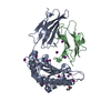 7p3eC  7pbeC 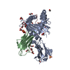 4i4wS C: citing same article ( S: Starting model for refinement |
|---|---|
| Similar structure data |


-Links
 PDBj
PDBj


- Assembly
| Deposited unit | 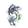
| ||||||||
|---|---|---|---|---|---|---|---|---|---|
| 1 |
| ||||||||
| Unit cell |
|
-Components
-Protein , 2 types, 2 molecules AB
| #1: Protein | Mass: 31951.316 Da / Num. of mol.: 1 Source method: isolated from a genetically manipulated source Source: (gene. exp.) Homo sapiens (human) / Gene: HLA-A / Production host:  Escherichia coli (E. coli) / References: UniProt: A0A5B8RNS7 Escherichia coli (E. coli) / References: UniProt: A0A5B8RNS7 |
|---|---|
| #2: Protein | Beta-2 microglobulin Mass: 11879.356 Da / Num. of mol.: 1 Source method: isolated from a genetically manipulated source Source: (gene. exp.) Homo sapiens (human) / Gene: B2M, CDABP0092, HDCMA22P / Production host: Escherichia coli (E. coli) / References: UniProt: P61769 |
-Protein/peptide , 1 types, 1 molecules C
| #3: Protein/peptide | Spike protein / S glycoprotein / E2 / Peplomer protein Mass: 1151.377 Da / Num. of mol.: 1 / Source method: obtained synthetically Source: (synth.) Severe acute respiratory syndrome coronavirus 2References: UniProt: P0DTC2 |
|---|
-Non-polymers , 4 types, 457 molecules 


| #4: Chemical | ChemComp-EDO / Ethylene glycol Mass: 62.068 Da / Num. of mol.: 6 / Source method: obtained synthetically / Formula: C2H6O2 Mass: 62.068 Da / Num. of mol.: 6 / Source method: obtained synthetically / Formula: C2H6O2#5: Chemical | ChemComp-ACT / | Acetate Mass: 59.044 Da / Num. of mol.: 1 / Source method: obtained synthetically / Formula: C2H3O2 Mass: 59.044 Da / Num. of mol.: 1 / Source method: obtained synthetically / Formula: C2H3O2#6: Chemical | ChemComp-CA / |  Mass: 40.078 Da / Num. of mol.: 1 / Source method: obtained synthetically / Formula: Ca Mass: 40.078 Da / Num. of mol.: 1 / Source method: obtained synthetically / Formula: Ca#7: Water | ChemComp-HOH / | WaterMass: 18.015 Da / Num. of mol.: 449 / Source method: isolated from a natural source / Formula: H2O |
|---|
-Details
| Has ligand of interest | N |
|---|
-Experimental details
-Experiment
| Experiment | Method: X-RAY DIFFRACTION / Number of used crystals: 1 |
|---|
- Sample preparation
| Crystal | Density Matthews: 2.58 Å3/Da / Density % sol: 52.41 % |
|---|---|
| Crystal grow | Temperature: 293 K / Method: vapor diffusion, sitting drop Details: 0.2 M sodium nitrate, 0.1 M Bis Tris propane pH 7.5, and 20 % w/v PEG 3350 Temp details: 7.5 |
-Data collection
| Diffraction | Mean temperature: 100 K / Serial crystal experiment: N |
|---|---|
| Diffraction source | Source: SYNCHROTRON / Site: Diamond  / Beamline: I04-1 / Wavelength: 0.91808 Å / Beamline: I04-1 / Wavelength: 0.91808 Å |
| Detector | Type: DECTRIS PILATUS 2M / Detector: PIXEL / Date: Jun 28, 2021 |
| Radiation | Protocol: SINGLE WAVELENGTH / Monochromatic (M) / Laue (L): M / Scattering type: x-ray |
| Radiation wavelength | Wavelength: 0.91808 Å / Relative weight: 1 |
| Reflection | Resolution: 1.67→79.39 Å / Num. obs: 53176 / % possible obs: 99.9 % / Redundancy: 3.9 % / CC1/2: 0.99 / Rmerge(I) obs: 0.162 / Rpim(I) all: 0.095 / Rrim(I) all: 0.188 / Net I/σ(I): 9.5 |
| Reflection shell | Resolution: 1.67→1.7 Å / Redundancy: 3.9 % / Rmerge(I) obs: 1.419 / Mean I/σ(I) obs: 0.9 / Num. unique obs: 2648 / CC1/2: 0.364 / Rpim(I) all: 0.839 / Rrim(I) all: 1.656 / % possible all: 100 |
-Phasing
| Phasing | Method: molecular replacement | |||||||||
|---|---|---|---|---|---|---|---|---|---|---|
| Phasing MR | Model details: Phaser MODE: MR_AUTO
|
- Processing
| Software |
| ||||||||||||||||||||||||||||||||||||||||||||||||||||||||||||||||||||||||||||||||||||||||||||||||||||
|---|---|---|---|---|---|---|---|---|---|---|---|---|---|---|---|---|---|---|---|---|---|---|---|---|---|---|---|---|---|---|---|---|---|---|---|---|---|---|---|---|---|---|---|---|---|---|---|---|---|---|---|---|---|---|---|---|---|---|---|---|---|---|---|---|---|---|---|---|---|---|---|---|---|---|---|---|---|---|---|---|---|---|---|---|---|---|---|---|---|---|---|---|---|---|---|---|---|---|---|---|---|
| Refinement | Method to determine structure: MOLECULAR REPLACEMENT Starting model: 4I4W Resolution: 1.67→51.89 Å / Cor.coef. Fo:Fc: 0.958 / Cor.coef. Fo:Fc free: 0.939 / SU B: 6.474 / SU ML: 0.103 / SU R Cruickshank DPI: 0.1043 / Cross valid method: THROUGHOUT / σ(F): 0 / ESU R: 0.104 / ESU R Free: 0.107 / Stereochemistry target values: MAXIMUM LIKELIHOOD Details: HYDROGENS HAVE BEEN ADDED IN THE RIDING POSITIONS U VALUES : WITH TLS ADDED
| ||||||||||||||||||||||||||||||||||||||||||||||||||||||||||||||||||||||||||||||||||||||||||||||||||||
| Solvent computation | Ion probe radii: 0.8 Å / Shrinkage radii: 0.8 Å / VDW probe radii: 1.2 Å / Solvent model: MASK | ||||||||||||||||||||||||||||||||||||||||||||||||||||||||||||||||||||||||||||||||||||||||||||||||||||
| Displacement parameters | Biso max: 85.44 Å2 / Biso mean: 20.099 Å2 / Biso min: 9.79 Å2
| ||||||||||||||||||||||||||||||||||||||||||||||||||||||||||||||||||||||||||||||||||||||||||||||||||||
| Refinement step | Cycle: final / Resolution: 1.67→51.89 Å
| ||||||||||||||||||||||||||||||||||||||||||||||||||||||||||||||||||||||||||||||||||||||||||||||||||||
| Refine LS restraints |
| ||||||||||||||||||||||||||||||||||||||||||||||||||||||||||||||||||||||||||||||||||||||||||||||||||||
| LS refinement shell | Resolution: 1.67→1.713 Å / Rfactor Rfree error: 0 / Total num. of bins used: 20
| ||||||||||||||||||||||||||||||||||||||||||||||||||||||||||||||||||||||||||||||||||||||||||||||||||||
| Refinement TLS params. | Method: refined / Refine-ID: X-RAY DIFFRACTION
| ||||||||||||||||||||||||||||||||||||||||||||||||||||||||||||||||||||||||||||||||||||||||||||||||||||
| Refinement TLS group |
|
