 Movie
Movie Controller

[English] 日本語
 Yorodumi
- PDB-7p2z: Crystal structure of human lysosomal acid-alpha-glucosidase, GAA,... -
+ Open data

- Basic information
| Entry | Database: PDB / ID: 7p2z | ||||||
|---|---|---|---|---|---|---|---|
| Title | Crystal structure of human lysosomal acid-alpha-glucosidase, GAA, in complex with cyclosulfamidate 4 | ||||||
 Components Components | Lysosomal alpha-glucosidase | ||||||
 Keywords Keywords |  HYDROLASE / alpha-glycosidase / lysosomal / Pompe disease / pharmacological chaperone HYDROLASE / alpha-glycosidase / lysosomal / Pompe disease / pharmacological chaperone | ||||||
| Function / homology |  Function and homology information Function and homology informationvacuolar sequestering / autolysosome lumen / maltose metabolic process / alpha-glucosidase activity / alpha-1,4-glucosidase activity / sucrose metabolic process / Glycogen storage disease type II (GAA) / glycophagy / alpha-glucosidase / maltose alpha-glucosidase activity ...vacuolar sequestering / autolysosome lumen / maltose metabolic process / alpha-glucosidase activity / alpha-1,4-glucosidase activity / sucrose metabolic process / Glycogen storage disease type II (GAA) / glycophagy / alpha-glucosidase / maltose alpha-glucosidase activity / diaphragm contraction / neuromuscular process controlling posture / tissue development / regulation of the force of heart contraction / glycogen catabolic process / aorta development / azurophil granule membrane / Glycogen breakdown (glycogenolysis) / muscle cell cellular homeostasis / lysosome organization / tertiary granule membrane / neuromuscular process controlling balance / ficolin-1-rich granule membrane / heart morphogenesis / cardiac muscle contraction / lysosomal lumen / locomotory behavior / glucose metabolic process / carbohydrate binding / lysosome / lysosomal membrane / intracellular membrane-bounded organelle / Neutrophil degranulation / extracellular exosome / membrane / plasma membraneSimilarity search - Function | ||||||
| Biological species |  Homo sapiens (human) Homo sapiens (human) | ||||||
| Method | X-RAY DIFFRACTION / SYNCHROTRON / FOURIER SYNTHESIS / Resolution: 1.85 Å | ||||||
 Authors Authors | Roig-Zamboni, V. / Kok, K. / Overkleeft, H. / Artola, M. / Sulzenbacher, G. | ||||||
 Citation Citation | Journal: J.Am.Chem.Soc. / Year: 2022 Title: 1,6- epi-Cyclophellitol Cyclosulfamidate Is a Bona Fide Lysosomal alpha-Glucosidase Stabilizer for the Treatment of Pompe Disease. Authors: Kok, K. / Kuo, C.L. / Katzy, R.E. / Lelieveld, L.T. / Wu, L. / Roig-Zamboni, V. / van der Marel, G.A. / Codee, J.D.C. / Sulzenbacher, G. / Davies, G.J. / Overkleeft, H.S. / Aerts, J.M.F.G. / Artola, M. | ||||||
| History |
|
- Structure visualization
| Structure viewer | Molecule: MolmilJmol/JSmol |
|---|
- Downloads & links
-Download
| PDBx/mmCIF format | 7p2z.cif.gz | 238.3 KB | Display | PDBx/mmCIF format |
|---|---|---|---|---|
| PDB format | pdb7p2z.ent.gz | Display | PDB format | |
| PDBx/mmJSON format | 7p2z.json.gz | Tree view | PDBx/mmJSON format | |
| Others |  Other downloads Other downloads |
-Validation report
| Arichive directory | https://data.pdbj.org/pub/pdb/validation_reports/p2/7p2zftp://data.pdbj.org/pub/pdb/validation_reports/p2/7p2z | HTTPS FTP |
|---|
-Related structure data
| Related structure data | 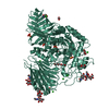 7p32C  7p4cC  7p4dC 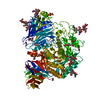 5nn4S S: Starting model for refinement C: citing same article ( |
|---|---|
| Similar structure data |
-Links
 PDBj
PDBj


- Assembly
| Deposited unit | 
| ||||||||
|---|---|---|---|---|---|---|---|---|---|
| 1 |
| ||||||||
| Unit cell |
|
-Components
-Protein , 1 types, 2 molecules AAAAaA
| #1: Protein | Mass: 96978.633 Da / Num. of mol.: 2 Source method: isolated from a genetically manipulated source Details: sample source is Myozyme / Source: (gene. exp.) Homo sapiens (human) / Gene: GAA / Cell line (production host): OVARY CELLS / Production host:   Cricetulus griseus (Chinese hamster) / References: UniProt: P10253, alpha-glucosidase Cricetulus griseus (Chinese hamster) / References: UniProt: P10253, alpha-glucosidase |
|---|
-Sugars , 4 types, 5 molecules
| #2: Polysaccharide | 2-acetamido-2-deoxy-beta-D-glucopyranose-(1-4)-[alpha-L-fucopyranose-(1-6)]2-acetamido-2-deoxy-beta- ...2-acetamido-2-deoxy-beta-D-glucopyranose-(1-4)-[alpha-L-fucopyranose-(1-6)]2-acetamido-2-deoxy-beta-D-glucopyranose / Mass: 570.542 Da / Num. of mol.: 1 Source method: isolated from a genetically manipulated source | ||||
|---|---|---|---|---|---|
| #3: Polysaccharide | / Mass: 424.401 Da / Num. of mol.: 2 Source method: isolated from a genetically manipulated source #4: Polysaccharide | beta-D-mannopyranose-(1-4)-2-acetamido-2-deoxy-beta-D-glucopyranose-(1-4)-2-acetamido-2-deoxy-beta- ...beta-D-mannopyranose-(1-4)-2-acetamido-2-deoxy-beta-D-glucopyranose-(1-4)-2-acetamido-2-deoxy-beta-D-glucopyranose | / Mass: 586.542 Da / Num. of mol.: 1Source method: isolated from a genetically manipulated source #5: Polysaccharide | alpha-L-fucopyranose-(1-6)-2-acetamido-2-deoxy-beta-D-glucopyranose | / Mass: 367.349 Da / Num. of mol.: 1Source method: isolated from a genetically manipulated source |
-Non-polymers , 7 types, 803 molecules 


| #6: Chemical | ChemComp-SO4 / Sulfate Mass: 96.063 Da / Num. of mol.: 5 / Source method: obtained synthetically / Formula: SO4 / Feature type: SUBJECT OF INVESTIGATION Mass: 96.063 Da / Num. of mol.: 5 / Source method: obtained synthetically / Formula: SO4 / Feature type: SUBJECT OF INVESTIGATION#7: Chemical | ChemComp-CL / Chloride Mass: 35.453 Da / Num. of mol.: 9 / Source method: obtained synthetically / Formula: Cl / Feature type: SUBJECT OF INVESTIGATION Mass: 35.453 Da / Num. of mol.: 9 / Source method: obtained synthetically / Formula: Cl / Feature type: SUBJECT OF INVESTIGATION#8: Chemical | Glycerol Mass: 92.094 Da / Num. of mol.: 3 / Source method: obtained synthetically / Formula: C3H8O3 / Feature type: SUBJECT OF INVESTIGATION Mass: 92.094 Da / Num. of mol.: 3 / Source method: obtained synthetically / Formula: C3H8O3 / Feature type: SUBJECT OF INVESTIGATION#9: Chemical | ChemComp-EDO / Ethylene glycol Mass: 62.068 Da / Num. of mol.: 5 / Source method: obtained synthetically / Formula: C2H6O2 / Feature type: SUBJECT OF INVESTIGATION Mass: 62.068 Da / Num. of mol.: 5 / Source method: obtained synthetically / Formula: C2H6O2 / Feature type: SUBJECT OF INVESTIGATION#10: Chemical | ChemComp-YTW / ( |  Mass: 255.246 Da / Num. of mol.: 1 / Source method: obtained synthetically / Formula: C7H13NO7S / Feature type: SUBJECT OF INVESTIGATION Mass: 255.246 Da / Num. of mol.: 1 / Source method: obtained synthetically / Formula: C7H13NO7S / Feature type: SUBJECT OF INVESTIGATION#11: Chemical | Polyethylene glycol Mass: 150.173 Da / Num. of mol.: 2 / Source method: obtained synthetically / Formula: C6H14O4 / Feature type: SUBJECT OF INVESTIGATION Mass: 150.173 Da / Num. of mol.: 2 / Source method: obtained synthetically / Formula: C6H14O4 / Feature type: SUBJECT OF INVESTIGATION#12: Water | ChemComp-HOH / | WaterMass: 18.015 Da / Num. of mol.: 778 / Source method: isolated from a natural source / Formula: H2O |
|---|
-Details
| Has ligand of interest | Y |
|---|
-Experimental details
-Experiment
| Experiment | Method: X-RAY DIFFRACTION / Number of used crystals: 1 |
|---|
- Sample preparation
| Crystal | Density Matthews: 3.4 Å3/Da / Density % sol: 63 % |
|---|---|
| Crystal grow | Temperature: 293 K / Method: vapor diffusion, hanging drop / pH: 7 Details: 1.9 M AMMONIUM SULPHATE, 0.1 M HEPES REMARK 280 PH 7.0, 2% V/V PEG400 |
-Data collection
| Diffraction | Mean temperature: 100 K / Serial crystal experiment: N |
|---|---|
| Diffraction source | Source: SYNCHROTRON / Site: SOLEIL  / Beamline: PROXIMA 2 / Wavelength: 0.9801 Å / Beamline: PROXIMA 2 / Wavelength: 0.9801 Å |
| Detector | Type: DECTRIS EIGER X 9M / Detector: PIXEL / Date: Sep 21, 2019 |
| Radiation | Protocol: SINGLE WAVELENGTH / Monochromatic (M) / Laue (L): M / Scattering type: x-ray |
| Radiation wavelength | Wavelength: 0.9801 Å / Relative weight: 1 |
| Reflection | Resolution: 1.85→47.65 Å / Num. obs: 110461 / % possible obs: 100 % / Redundancy: 13.6 % / Biso Wilson estimate: 20.85 Å2 / CC1/2: 0.998 / Rmerge(I) obs: 0.121 / Rpim(I) all: 0.049 / Rrim(I) all: 0.13 / Net I/σ(I): 14.5 |
| Reflection shell | Resolution: 1.85→1.88 Å / Rmerge(I) obs: 1.554 / Mean I/σ(I) obs: 2.1 / Num. unique obs: 5401 / CC1/2: 0.742 / Rpim(I) all: 0.628 / % possible all: 99.9 |
- Processing
| Software |
| |||||||||||||||||||||||||||||||||||||||||||||||||||||||||||||||||||||||||||||||||||||||||||||||||||||||||||||||||||||||||||||||||||||||||||||||||||||||||||
|---|---|---|---|---|---|---|---|---|---|---|---|---|---|---|---|---|---|---|---|---|---|---|---|---|---|---|---|---|---|---|---|---|---|---|---|---|---|---|---|---|---|---|---|---|---|---|---|---|---|---|---|---|---|---|---|---|---|---|---|---|---|---|---|---|---|---|---|---|---|---|---|---|---|---|---|---|---|---|---|---|---|---|---|---|---|---|---|---|---|---|---|---|---|---|---|---|---|---|---|---|---|---|---|---|---|---|---|---|---|---|---|---|---|---|---|---|---|---|---|---|---|---|---|---|---|---|---|---|---|---|---|---|---|---|---|---|---|---|---|---|---|---|---|---|---|---|---|---|---|---|---|---|---|---|---|---|
| Refinement | Method to determine structure: FOURIER SYNTHESIS Starting model: 5nn4 Resolution: 1.85→47 Å / Cor.coef. Fo:Fc: 0.975 / Cor.coef. Fo:Fc free: 0.966 / SU B: 2.132 / SU ML: 0.062 / Cross valid method: FREE R-VALUE / ESU R: 0.091 / ESU R Free: 0.09 Details: Hydrogens have been added in their riding positions
| |||||||||||||||||||||||||||||||||||||||||||||||||||||||||||||||||||||||||||||||||||||||||||||||||||||||||||||||||||||||||||||||||||||||||||||||||||||||||||
| Solvent computation | Ion probe radii: 0.7 Å / Shrinkage radii: 0.7 Å / VDW probe radii: 1.1 Å / Solvent model: MASK BULK SOLVENT | |||||||||||||||||||||||||||||||||||||||||||||||||||||||||||||||||||||||||||||||||||||||||||||||||||||||||||||||||||||||||||||||||||||||||||||||||||||||||||
| Displacement parameters | Biso mean: 28.703 Å2
| |||||||||||||||||||||||||||||||||||||||||||||||||||||||||||||||||||||||||||||||||||||||||||||||||||||||||||||||||||||||||||||||||||||||||||||||||||||||||||
| Refinement step | Cycle: LAST / Resolution: 1.85→47 Å
| |||||||||||||||||||||||||||||||||||||||||||||||||||||||||||||||||||||||||||||||||||||||||||||||||||||||||||||||||||||||||||||||||||||||||||||||||||||||||||
| Refine LS restraints |
| |||||||||||||||||||||||||||||||||||||||||||||||||||||||||||||||||||||||||||||||||||||||||||||||||||||||||||||||||||||||||||||||||||||||||||||||||||||||||||
| LS refinement shell |
|
