 Movie
Movie Controller

[English] 日本語
 Yorodumi
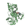
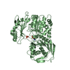
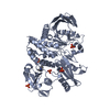
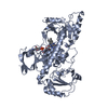
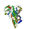
- PDB-7ouj: Crystal structure of the flavoprotein monooxygenase RubL from rub... -
+ Open data

- Basic information
| Entry | Database: PDB / ID: 7ouj | ||||||||||||
|---|---|---|---|---|---|---|---|---|---|---|---|---|---|
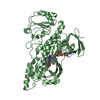
| Title | Crystal structure of the flavoprotein monooxygenase RubL from rubromycin biosynthesis | ||||||||||||
 Components Components | RubL | ||||||||||||
 Keywords Keywords |  FLAVOPROTEIN / monooxygenase / griseorhodin A biosynthesis / rubromycin biosynthesis FLAVOPROTEIN / monooxygenase / griseorhodin A biosynthesis / rubromycin biosynthesis | ||||||||||||
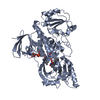
| Function / homology | FAD-binding domain / FAD binding domain / FAD binding / FAD/NAD(P)-binding domain superfamily / (2S)-hexane-1,2,6-triol / FLAVIN-ADENINE DINUCLEOTIDE / 4-HYDROXYPROLINE / Putative polyketide hydroxylase Function and homology information Function and homology information | ||||||||||||
| Biological species |  Streptomyces collinus (bacteria) Streptomyces collinus (bacteria) | ||||||||||||
| Method | X-RAY DIFFRACTION / SYNCHROTRON / MOLECULAR REPLACEMENT / Resolution: 1.573 Å | ||||||||||||
 Authors Authors | Saleem-Batcha, R. / Toplak, M. / Teufel, R. | ||||||||||||
| Funding support |  Germany, Germany,  Austria, 3items Austria, 3items
| ||||||||||||
 Citation Citation | Journal: Angew.Chem.Int.Ed.Engl. / Year: 2021 Title: Catalytic Control of Spiroketal Formation in Rubromycin Polyketide Biosynthesis. Authors: Toplak, M. / Saleem-Batcha, R. / Piel, J. / Teufel, R. | ||||||||||||
| History |
|
- Structure visualization
| Structure viewer | Molecule: MolmilJmol/JSmol |
|---|
- Downloads & links
-Download
| PDBx/mmCIF format | 7ouj.cif.gz | 125.2 KB | Display | PDBx/mmCIF format |
|---|---|---|---|---|
| PDB format | pdb7ouj.ent.gz | Display | PDB format | |
| PDBx/mmJSON format | 7ouj.json.gz | Tree view | PDBx/mmJSON format | |
| Others |  Other downloads Other downloads |
-Validation report
| Arichive directory | https://data.pdbj.org/pub/pdb/validation_reports/ou/7oujftp://data.pdbj.org/pub/pdb/validation_reports/ou/7ouj | HTTPS FTP |
|---|
-Related structure data
| Related structure data | 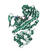 7oucC  7oudC 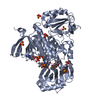 3ihgS C: citing same article ( S: Starting model for refinement |
|---|---|
| Similar structure data |


-Links
 PDBj
PDBj- Assembly
| Deposited unit | 
| ||||||||
|---|---|---|---|---|---|---|---|---|---|
| 1 |
| ||||||||
| Unit cell |
|
-Components
-Protein , 1 types, 1 molecules AAA
| #1: Protein | Mass: 57918.562 Da / Num. of mol.: 1 Source method: isolated from a genetically manipulated source Details: HYP is ligand from crystallisation buffer / Source: (gene. exp.) Streptomyces collinus (bacteria) / Gene: rubL / Production host: Escherichia coli BL21(DE3) (bacteria) / References: UniProt: Q8KY42 |
|---|
-Non-polymers , 6 types, 201 molecules 


| #2: Chemical | ChemComp-FAD / Flavin adenine dinucleotide Mass: 785.550 Da / Num. of mol.: 1 / Source method: obtained synthetically / Formula: C27H33N9O15P2 / Feature type: SUBJECT OF INVESTIGATION / Comment: FAD*YM Mass: 785.550 Da / Num. of mol.: 1 / Source method: obtained synthetically / Formula: C27H33N9O15P2 / Feature type: SUBJECT OF INVESTIGATION / Comment: FAD*YM | ||||||
|---|---|---|---|---|---|---|---|
| #3: Chemical | ChemComp-CL / Chloride Mass: 35.453 Da / Num. of mol.: 1 / Source method: obtained synthetically / Formula: Cl / Feature type: SUBJECT OF INVESTIGATION Mass: 35.453 Da / Num. of mol.: 1 / Source method: obtained synthetically / Formula: Cl / Feature type: SUBJECT OF INVESTIGATION | ||||||
| #4: Chemical |  Mass: 134.174 Da / Num. of mol.: 3 / Source method: obtained synthetically / Formula: C6H14O3 Mass: 134.174 Da / Num. of mol.: 3 / Source method: obtained synthetically / Formula: C6H14O3#5: Chemical | ChemComp-HYP / | Hydroxyproline Type: L-peptide linking / Mass: 131.130 Da / Num. of mol.: 1 / Source method: obtained synthetically / Formula: C5H9NO3 Type: L-peptide linking / Mass: 131.130 Da / Num. of mol.: 1 / Source method: obtained synthetically / Formula: C5H9NO3#6: Chemical | ChemComp-GOL / | Glycerol Mass: 92.094 Da / Num. of mol.: 1 / Source method: isolated from a natural source / Formula: C3H8O3 Mass: 92.094 Da / Num. of mol.: 1 / Source method: isolated from a natural source / Formula: C3H8O3#7: Water | ChemComp-HOH / | WaterMass: 18.015 Da / Num. of mol.: 194 / Source method: isolated from a natural source / Formula: H2O |
-Details
| Has ligand of interest | Y |
|---|
-Experimental details
-Experiment
| Experiment | Method: X-RAY DIFFRACTION / Number of used crystals: 1 |
|---|
- Sample preparation
| Crystal | Density Matthews: 2.34 Å3/Da / Density % sol: 47.43 % |
|---|---|
| Crystal grow | Temperature: 295 K / Method: vapor diffusion, sitting drop / pH: 7.5 Details: 20 mM DL-Arginine hydrochloride, 20 mM DL-Threonine, 20 mM DL-Histidine monohydrochloride monohydrate, 20 mM DL-5-Hydroxylysine hydrochloride, 20 mM trans-4-hydroxy-L-proline, 100 mM BES/TEA ...Details: 20 mM DL-Arginine hydrochloride, 20 mM DL-Threonine, 20 mM DL-Histidine monohydrochloride monohydrate, 20 mM DL-5-Hydroxylysine hydrochloride, 20 mM trans-4-hydroxy-L-proline, 100 mM BES/TEA pH 7.5 buffer system, 12.5% PEG 4000 and 20% 1,2,6-Hexanetriol |
-Data collection
| Diffraction | Mean temperature: 100 K / Serial crystal experiment: N |
|---|---|
| Diffraction source | Source: SYNCHROTRON / Site: SLS  / Beamline: X06SA / Wavelength: 1 Å / Beamline: X06SA / Wavelength: 1 Å |
| Detector | Type: DECTRIS EIGER X 16M / Detector: PIXEL / Date: Jul 9, 2020 |
| Radiation | Monochromator: M / Protocol: SINGLE WAVELENGTH / Monochromatic (M) / Laue (L): M / Scattering type: x-ray |
| Radiation wavelength | Wavelength: 1 Å / Relative weight: 1 |
| Reflection | Resolution: 1.57→46.08 Å / Num. obs: 70014 / % possible obs: 94 % / Redundancy: 2.4 % / CC1/2: 0.99 / Rmerge(I) obs: 0.046 / Net I/σ(I): 9.4 |
| Reflection shell | Resolution: 1.57→1.66 Å / Rmerge(I) obs: 0.55 / Num. unique obs: 10481 / CC1/2: 0.7 |
- Processing
| Software |
| |||||||||||||||||||||||||||||||||||||||||||||||||||||||||||||||||||||||||||||||||||||||||||||||||||||||||||||||||||||||||||||||||||||||||||||||||||||||||||
|---|---|---|---|---|---|---|---|---|---|---|---|---|---|---|---|---|---|---|---|---|---|---|---|---|---|---|---|---|---|---|---|---|---|---|---|---|---|---|---|---|---|---|---|---|---|---|---|---|---|---|---|---|---|---|---|---|---|---|---|---|---|---|---|---|---|---|---|---|---|---|---|---|---|---|---|---|---|---|---|---|---|---|---|---|---|---|---|---|---|---|---|---|---|---|---|---|---|---|---|---|---|---|---|---|---|---|---|---|---|---|---|---|---|---|---|---|---|---|---|---|---|---|---|---|---|---|---|---|---|---|---|---|---|---|---|---|---|---|---|---|---|---|---|---|---|---|---|---|---|---|---|---|---|---|---|---|
| Refinement | Method to determine structure: MOLECULAR REPLACEMENT Starting model: 3IHG Resolution: 1.573→37.778 Å / Cor.coef. Fo:Fc: 0.969 / Cor.coef. Fo:Fc free: 0.96 / WRfactor Rfree: 0.214 / WRfactor Rwork: 0.18 / SU B: 2.014 / SU ML: 0.066 / Average fsc free: 0.8812 / Average fsc work: 0.8859 / Cross valid method: FREE R-VALUE / ESU R: 0.088 / ESU R Free: 0.087 Details: Hydrogens have been added in their riding positions
| |||||||||||||||||||||||||||||||||||||||||||||||||||||||||||||||||||||||||||||||||||||||||||||||||||||||||||||||||||||||||||||||||||||||||||||||||||||||||||
| Solvent computation | Ion probe radii: 0.8 Å / Shrinkage radii: 0.8 Å / VDW probe radii: 1.2 Å / Solvent model: MASK BULK SOLVENT | |||||||||||||||||||||||||||||||||||||||||||||||||||||||||||||||||||||||||||||||||||||||||||||||||||||||||||||||||||||||||||||||||||||||||||||||||||||||||||
| Displacement parameters | Biso mean: 27.196 Å2
| |||||||||||||||||||||||||||||||||||||||||||||||||||||||||||||||||||||||||||||||||||||||||||||||||||||||||||||||||||||||||||||||||||||||||||||||||||||||||||
| Refinement step | Cycle: LAST / Resolution: 1.573→37.778 Å
| |||||||||||||||||||||||||||||||||||||||||||||||||||||||||||||||||||||||||||||||||||||||||||||||||||||||||||||||||||||||||||||||||||||||||||||||||||||||||||
| Refine LS restraints |
| |||||||||||||||||||||||||||||||||||||||||||||||||||||||||||||||||||||||||||||||||||||||||||||||||||||||||||||||||||||||||||||||||||||||||||||||||||||||||||
| LS refinement shell |
|
