 Movie
Movie Controller

[English] 日本語
 Yorodumi
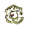
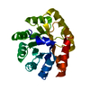
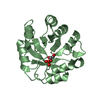
- PDB-7osv: DeNovoTIM6-SB, a de novo designed TIM barrel with a salt-bridge c... -
+ Open data

- Basic information
| Entry | Database: PDB / ID: 7osv | |||||||||
|---|---|---|---|---|---|---|---|---|---|---|
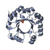
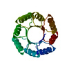
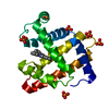
| Title | DeNovoTIM6-SB, a de novo designed TIM barrel with a salt-bridge cluster (crystal form 1) | |||||||||
 Components Components | DeNovoTIM6-SB | |||||||||
 Keywords Keywords |  DE NOVO PROTEIN / TIM barrel / salt bridge cluster DE NOVO PROTEIN / TIM barrel / salt bridge cluster | |||||||||
| Function / homology | ACETATE ION / DI(HYDROXYETHYL)ETHER Function and homology information Function and homology information | |||||||||
| Biological species | synthetic construct (others) | |||||||||
| Method | X-RAY DIFFRACTION / SYNCHROTRON / MOLECULAR REPLACEMENT / Resolution: 1.66 Å | |||||||||
 Authors Authors | Kordes, S. / Romero-Romero, S. / Hocker, B. | |||||||||
| Funding support |  Germany, 2items Germany, 2items
| |||||||||
 Citation Citation | Journal: Protein Sci. / Year: 2022 Title: A newly introduced salt bridge cluster improves structural and biophysical properties of de novo TIM barrels. Authors: Kordes, S. / Romero-Romero, S. / Lutz, L. / Hocker, B. | |||||||||
| History |
|
- Structure visualization
| Structure viewer | Molecule: MolmilJmol/JSmol |
|---|
- Downloads & links
-Download
| PDBx/mmCIF format | 7osv.cif.gz | 136.4 KB | Display | PDBx/mmCIF format |
|---|---|---|---|---|
| PDB format | pdb7osv.ent.gz | 89.7 KB | Display | PDB format |
| PDBx/mmJSON format | 7osv.json.gz | Tree view | PDBx/mmJSON format | |
| Others |  Other downloads Other downloads |
-Validation report
| Arichive directory | https://data.pdbj.org/pub/pdb/validation_reports/os/7osvftp://data.pdbj.org/pub/pdb/validation_reports/os/7osv | HTTPS FTP |
|---|
-Related structure data
| Related structure data |  7osuC  7ot7C  7ot8C 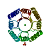 7p12C  5bvlS S: Starting model for refinement C: citing same article ( |
|---|---|
| Similar structure data |


-Links
 PDBj
PDBj
- Assembly
| Deposited unit | 
| ||||||||||||
|---|---|---|---|---|---|---|---|---|---|---|---|---|---|
| 1 |
| ||||||||||||
| Unit cell |
|
-Components
| #1: Protein | Mass: 22669.711 Da / Num. of mol.: 1 Source method: isolated from a genetically manipulated source Details: M (N-terminal) and GLEHHHHHH (C-terminal) come from the expresion vector. Source: (gene. exp.) synthetic construct (others) / Plasmid: pET21b(+) / Production host:  Escherichia coli BL21(DE3) (bacteria) Escherichia coli BL21(DE3) (bacteria) | ||||||
|---|---|---|---|---|---|---|---|
| #2: Chemical | ChemComp-SO4 / Sulfate  Mass: 96.063 Da / Num. of mol.: 1 / Source method: obtained synthetically / Formula: SO4 Mass: 96.063 Da / Num. of mol.: 1 / Source method: obtained synthetically / Formula: SO4 | ||||||
| #3: Chemical | Acetate  Mass: 59.044 Da / Num. of mol.: 2 / Source method: obtained synthetically / Formula: C2H3O2 Mass: 59.044 Da / Num. of mol.: 2 / Source method: obtained synthetically / Formula: C2H3O2#4: Chemical | ChemComp-PEG / | Diethylene glycol  Mass: 106.120 Da / Num. of mol.: 1 / Source method: obtained synthetically / Formula: C4H10O3 Mass: 106.120 Da / Num. of mol.: 1 / Source method: obtained synthetically / Formula: C4H10O3#5: Water | ChemComp-HOH / | Water Mass: 18.015 Da / Num. of mol.: 115 / Source method: isolated from a natural source / Formula: H2O Mass: 18.015 Da / Num. of mol.: 115 / Source method: isolated from a natural source / Formula: H2OHas ligand of interest | N | |
-Experimental details
-Experiment
| Experiment | Method: X-RAY DIFFRACTION / Number of used crystals: 1 |
|---|
- Sample preparation
| Crystal | Density Matthews: 1.96 Å3/Da / Density % sol: 37.3 % |
|---|---|
| Crystal grow | Temperature: 293.15 K / Method: vapor diffusion, sitting drop / pH: 4.6 Details: 0.2 M Ammonium sulfate, 0.2 M sodium acetate pH 4.6, 28% PEG 4000 |
-Data collection
| Diffraction | Mean temperature: 100 K / Serial crystal experiment: N |
|---|---|
| Diffraction source | Source: SYNCHROTRON / Site: SLS  / Beamline: X06SA / Wavelength: 1 Å / Beamline: X06SA / Wavelength: 1 Å |
| Detector | Type: DECTRIS EIGER X 16M / Detector: PIXEL / Date: Dec 16, 2020 |
| Radiation | Protocol: SINGLE WAVELENGTH / Monochromatic (M) / Laue (L): M / Scattering type: x-ray |
| Radiation wavelength | Wavelength: 1 Å / Relative weight: 1 |
| Reflection | Resolution: 1.66→32.81 Å / Num. obs: 19751 / % possible obs: 98.89 % / Redundancy: 6.9 % / Biso Wilson estimate: 27.41 Å2 / CC1/2: 0.997 / CC star: 0.999 / Rmerge(I) obs: 0.1385 / Rpim(I) all: 0.057 / Rrim(I) all: 0.1501 / Net I/σ(I): 7.77 |
| Reflection shell | Resolution: 1.66→1.72 Å / Redundancy: 7 % / Rmerge(I) obs: 1.77 / Mean I/σ(I) obs: 0.96 / Num. unique obs: 1947 / CC1/2: 0.339 / CC star: 0.712 / Rpim(I) all: 1.114 / Rrim(I) all: 2.99 / % possible all: 97.45 |
- Processing
| Software |
| ||||||||||||||||||||||||||||||||||||||||||||||||||||||||
|---|---|---|---|---|---|---|---|---|---|---|---|---|---|---|---|---|---|---|---|---|---|---|---|---|---|---|---|---|---|---|---|---|---|---|---|---|---|---|---|---|---|---|---|---|---|---|---|---|---|---|---|---|---|---|---|---|---|
| Refinement | Method to determine structure: MOLECULAR REPLACEMENT Starting model: 5BVL Resolution: 1.66→32.81 Å / SU ML: 0.2675 / Cross valid method: FREE R-VALUE / σ(F): 1.36 / Phase error: 29.6892 Stereochemistry target values: GeoStd + Monomer Library + CDL v1.2
| ||||||||||||||||||||||||||||||||||||||||||||||||||||||||
| Solvent computation | Shrinkage radii: 0.9 Å / VDW probe radii: 1.11 Å / Solvent model: FLAT BULK SOLVENT MODEL | ||||||||||||||||||||||||||||||||||||||||||||||||||||||||
| Displacement parameters | Biso mean: 33.7 Å2 | ||||||||||||||||||||||||||||||||||||||||||||||||||||||||
| Refinement step | Cycle: LAST / Resolution: 1.66→32.81 Å
| ||||||||||||||||||||||||||||||||||||||||||||||||||||||||
| Refine LS restraints |
| ||||||||||||||||||||||||||||||||||||||||||||||||||||||||
| LS refinement shell |
| ||||||||||||||||||||||||||||||||||||||||||||||||||||||||
| Refinement TLS params. | Method: refined / Origin x: 17.9931368244 Å / Origin y: 23.1375605104 Å / Origin z: 32.2013283573 Å
| ||||||||||||||||||||||||||||||||||||||||||||||||||||||||
| Refinement TLS group | Selection details: all |
