 Movie
Movie Controller

[English] 日本語
 Yorodumi
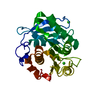
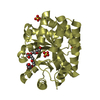
- PDB-7osb: Crystal Structure of a Double Mutant PETase (S238F/W159H) from Id... -
+ Open data

- Basic information
| Entry | Database: PDB / ID: 7osb | |||||||||
|---|---|---|---|---|---|---|---|---|---|---|
| Title | Crystal Structure of a Double Mutant PETase (S238F/W159H) from Ideonella sakaiensis | |||||||||
 Components Components | Poly(ethylene terephthalate) hydrolase | |||||||||
 Keywords Keywords |  HYDROLASE HYDROLASE | |||||||||
| Function / homology |  Function and homology information Function and homology informationpoly(ethylene terephthalate) hydrolase / acetylesterase activity / carboxylic ester hydrolase activity / : / xenobiotic catabolic process / extracellular regionSimilarity search - Function | |||||||||
| Biological species |  Ideonella sakaiensis (bacteria) Ideonella sakaiensis (bacteria) | |||||||||
| Method | X-RAY DIFFRACTION / SYNCHROTRON / MOLECULAR REPLACEMENT / Resolution: 1.45 Å | |||||||||
 Authors Authors | Shakespeare, T.J. / Zahn, M. / Allen, M.D. / McGeehan, J.E. | |||||||||
| Funding support |  United Kingdom, United Kingdom,  United States, 2items United States, 2items
| |||||||||
 Citation Citation | Journal: ChemSusChem / Year: 2022 Title: Comparative Performance of PETase as a Function of Reaction Conditions, Substrate Properties, and Product Accumulation. Authors: Erickson, E. / Shakespeare, T.J. / Bratti, F. / Buss, B.L. / Graham, R. / Hawkins, M.A. / Konig, G. / Michener, W.E. / Miscall, J. / Ramirez, K.J. / Rorrer, N.A. / Zahn, M. / Pickford, A.R. ...Authors: Erickson, E. / Shakespeare, T.J. / Bratti, F. / Buss, B.L. / Graham, R. / Hawkins, M.A. / Konig, G. / Michener, W.E. / Miscall, J. / Ramirez, K.J. / Rorrer, N.A. / Zahn, M. / Pickford, A.R. / McGeehan, J.E. / Beckham, G.T. | |||||||||
| History |
|
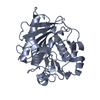
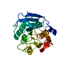
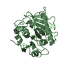
- Structure visualization
| Structure viewer | Molecule: MolmilJmol/JSmol |
|---|
- Downloads & links
-Download
| PDBx/mmCIF format | 7osb.cif.gz | 322.5 KB | Display | PDBx/mmCIF format |
|---|---|---|---|---|
| PDB format | pdb7osb.ent.gz | 262.3 KB | Display | PDB format |
| PDBx/mmJSON format | 7osb.json.gz | Tree view | PDBx/mmJSON format | |
| Others |  Other downloads Other downloads |
-Validation report
| Arichive directory | https://data.pdbj.org/pub/pdb/validation_reports/os/7osbftp://data.pdbj.org/pub/pdb/validation_reports/os/7osb | HTTPS FTP |
|---|
-Related structure data
| Related structure data | 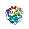 6eqeS S: Starting model for refinement |
|---|---|
| Similar structure data |


-Links
 PDBj
PDBj
- Assembly
| Deposited unit | 
| ||||||||
|---|---|---|---|---|---|---|---|---|---|
| 1 | 
| ||||||||
| 2 | 
| ||||||||
| 3 | 
| ||||||||
| Unit cell |
| ||||||||
| Components on special symmetry positions |
|
-Components
| #1: Protein | Mass: 31353.955 Da / Num. of mol.: 3 / Mutation: S238F, W159H Source method: isolated from a genetically manipulated source Source: (gene. exp.) Ideonella sakaiensis (bacteria) / Gene: ISF6_4831 / Production host: Escherichia coli (E. coli)References: UniProt: A0A0K8P6T7, poly(ethylene terephthalate) hydrolase #2: Chemical | ChemComp-SO4 / Sulfate  Mass: 96.063 Da / Num. of mol.: 10 / Source method: obtained synthetically / Formula: SO4 Mass: 96.063 Da / Num. of mol.: 10 / Source method: obtained synthetically / Formula: SO4#3: Chemical | Chloride  Mass: 35.453 Da / Num. of mol.: 3 / Source method: obtained synthetically / Formula: Cl Mass: 35.453 Da / Num. of mol.: 3 / Source method: obtained synthetically / Formula: Cl#4: Chemical | Glycerol  Mass: 92.094 Da / Num. of mol.: 2 / Source method: obtained synthetically / Formula: C3H8O3 Mass: 92.094 Da / Num. of mol.: 2 / Source method: obtained synthetically / Formula: C3H8O3#5: Water | ChemComp-HOH / | Water Mass: 18.015 Da / Num. of mol.: 1082 / Source method: isolated from a natural source / Formula: H2O Mass: 18.015 Da / Num. of mol.: 1082 / Source method: isolated from a natural source / Formula: H2OHas ligand of interest | N | |
|---|
-Experimental details
-Experiment
| Experiment | Method: X-RAY DIFFRACTION / Number of used crystals: 1 |
|---|
- Sample preparation
| Crystal | Density Matthews: 2.68 Å3/Da / Density % sol: 54.17 % |
|---|---|
| Crystal grow | Temperature: 293 K / Method: vapor diffusion, sitting drop / pH: 6.5 Details: 1.95M Ammonium Sulfate, 0.1M Sodium Citrate, pH 6.5 |
-Data collection
| Diffraction | Mean temperature: 100 K / Serial crystal experiment: N |
|---|---|
| Diffraction source | Source: SYNCHROTRON / Site: Diamond / Beamline: I03 / Wavelength: 0.9763 Å |
| Detector | Type: DECTRIS EIGER2 X 16M / Detector: PIXEL / Date: Aug 5, 2019 |
| Radiation | Protocol: SINGLE WAVELENGTH / Monochromatic (M) / Laue (L): M / Scattering type: x-ray |
| Radiation wavelength | Wavelength: 0.9763 Å / Relative weight: 1 |
| Reflection | Resolution: 1.45→20.7 Å / Num. obs: 178688 / % possible obs: 99.69 % / Redundancy: 1.99 % / Biso Wilson estimate: 18.11 Å2 / CC1/2: 0.999 / CC star: 1 / Rmerge(I) obs: 0.02805 / Rpim(I) all: 0.02805 / Rrim(I) all: 0.03966 / Net I/σ(I): 9.99 |
| Reflection shell | Resolution: 1.45→1.502 Å / Rmerge(I) obs: 0.6994 / Mean I/σ(I) obs: 1.11 / Num. unique obs: 17558 / CC1/2: 0.639 / CC star: 0.883 / Rpim(I) all: 0.6994 / Rrim(I) all: 0.9891 / % possible all: 98.83 |
- Processing
| Software |
| ||||||||||||||||||||||||||||||||||||||||||||||||||||||||||||||||||||||||||||||||||||||||||||||||||||
|---|---|---|---|---|---|---|---|---|---|---|---|---|---|---|---|---|---|---|---|---|---|---|---|---|---|---|---|---|---|---|---|---|---|---|---|---|---|---|---|---|---|---|---|---|---|---|---|---|---|---|---|---|---|---|---|---|---|---|---|---|---|---|---|---|---|---|---|---|---|---|---|---|---|---|---|---|---|---|---|---|---|---|---|---|---|---|---|---|---|---|---|---|---|---|---|---|---|---|---|---|---|
| Refinement | Method to determine structure: MOLECULAR REPLACEMENT Starting model: 6EQE Resolution: 1.45→21.02 Å / Cor.coef. Fo:Fc: 0.931 / Cor.coef. Fo:Fc free: 0.927 / SU R Cruickshank DPI: 0.06 / Cross valid method: THROUGHOUT / SU R Blow DPI: 0.065 / SU Rfree Blow DPI: 0.064 / SU Rfree Cruickshank DPI: 0.059
| ||||||||||||||||||||||||||||||||||||||||||||||||||||||||||||||||||||||||||||||||||||||||||||||||||||
| Displacement parameters | Biso mean: 30.28 Å2
| ||||||||||||||||||||||||||||||||||||||||||||||||||||||||||||||||||||||||||||||||||||||||||||||||||||
| Refine analyze | Luzzati coordinate error obs: 0.29 Å | ||||||||||||||||||||||||||||||||||||||||||||||||||||||||||||||||||||||||||||||||||||||||||||||||||||
| Refinement step | Cycle: LAST / Resolution: 1.45→21.02 Å
| ||||||||||||||||||||||||||||||||||||||||||||||||||||||||||||||||||||||||||||||||||||||||||||||||||||
| Refine LS restraints |
| ||||||||||||||||||||||||||||||||||||||||||||||||||||||||||||||||||||||||||||||||||||||||||||||||||||
| LS refinement shell | Resolution: 1.45→1.46 Å
| ||||||||||||||||||||||||||||||||||||||||||||||||||||||||||||||||||||||||||||||||||||||||||||||||||||
| Refinement TLS params. | Refine-ID: X-RAY DIFFRACTION
| ||||||||||||||||||||||||||||||||||||||||||||||||||||||||||||||||||||||||||||||||||||||||||||||||||||
| Refinement TLS group |
|
